 Back
Chapter 16: The Endocrine System – Anatomy & Physiology Study Notes
Study Guide - Smart Notes
 Tailored notes based on your materials, expanded with key definitions, examples, and context.
The Endocrine System
Overview of the Endocrine System
The endocrine system is one of the two major regulatory systems of the body, alongside the nervous system. It consists of organs that synthesize and secrete chemical messengers called hormones into the blood, which interact with specific cells known as target cells. Tissues containing these target cells are referred to as target tissues. Target cells possess receptors for hormones, enabling changes in cellular function.
Hormones are secreted into the interstitial fluid and then diffuse into blood capillaries.
The blood transports hormones throughout the body, allowing them to reach distant targets.
Hormones bind to receptors on target cells, initiating physiological responses.
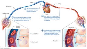
Comparison of the Endocrine and Nervous Systems
The endocrine system regulates body functions by releasing hormones into the bloodstream, whereas the nervous system uses electrical and chemical signals for rapid, short-term control.
Endocrine cells do not come into direct contact with target cells; hormones travel via blood.
Effects of hormones are generally slower but longer-lasting than nervous system signals.
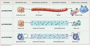
Paracrine and Autocrine Signals
Not all chemical messengers act as classic hormones. Some act locally:
Paracrine signals: Chemicals secreted by cells into the extracellular fluid (ECF) to influence nearby cells.
Autocrine signals: Chemicals secreted by a cell into the ECF to elicit effects from the same cell.
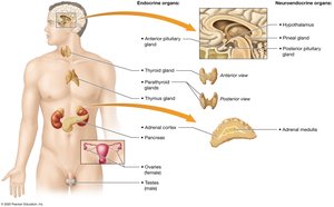
Overview of Endocrine Organs
Endocrine glands are ductless organs composed of glandular epithelial cells that secrete hormones into the interstitial fluid for transport by the bloodstream. This contrasts with exocrine glands, which secrete products into ducts leading to body surfaces or cavities.
Primary endocrine organs: anterior pituitary, thyroid, parathyroid, adrenal cortices, pancreas, thymus, ovaries/testes.
Secondary endocrine glands: heart, kidneys, small intestine, adipose tissue.
Neuroendocrine organs: hypothalamus, pineal gland, adrenal medulla.
Hormones: Structure, Transport, and Action
Classes of Hormones
Amino acid-based hormones: Derived from amino acids; mostly hydrophilic; produced in hypothalamus, adrenal medullae, thyroid gland.
Peptide/protein hormones: Chains of amino acids; generally hydrophilic; produced in hypothalamus, pancreas, anterior pituitary, thyroid, parathyroid.
Steroid hormones: Cholesterol derivatives; hydrophobic (lipid-soluble); produced in adrenal cortices, testes, ovaries.
Hormone Transport
Free hormones: Small, hydrophilic hormones travel freely in plasma.
Bound hormones: Hydrophobic hormones form complexes with binding proteins, extending their lifespan in blood.
Target Cells and Receptors
Target cells contain highly specific receptors for hormones, which can be located in the plasma membrane, cytosol, or nucleus.
Some hormones bind to one type of target cell; others bind to multiple cell types.
Hormones may bind to a single receptor or multiple receptors, producing different effects.
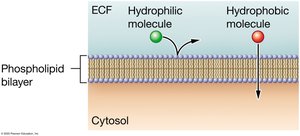
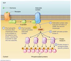
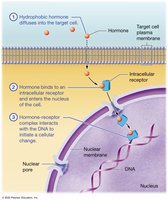
Receptors in the plasma membrane bind hydrophilic hormones; intracellular receptors bind hydrophobic hormones.
Regulation of Receptor Number
Upregulation: Target cells increase receptor number when hormone levels decline.
Downregulation: Target cells decrease receptor number after prolonged exposure to high hormone levels.
Mechanisms of Hormone Action
Hormones act via different mechanisms depending on their chemical nature:
Hydrophilic hormones: Bind to cell surface receptors and activate second-messenger systems (e.g., cAMP pathway).
Hydrophobic hormones: Diffuse into cells, bind to intracellular receptors, and directly influence gene expression.
Effects of Hormone Actions
Stimulate secretion from cells
Activate/inhibit enzymes
Stimulate/inhibit mitosis or meiosis
Open/close ion channels
Activate/inhibit gene transcription
Hormone Interactions
Complementary actions: Hormones affect different target cells to achieve a common goal.
Synergists: Hormones act on the same target cell to exert the same effect.
Antagonists: Hormones act on the same target cell but have opposite effects.
Hormone Half-Life and Elimination
Hormones are removed by kidneys (urine) or liver (enzymatic breakdown).
Half-life: Time for plasma concentration to reduce by half; hydrophobic hormones generally have longer half-lives.
Regulation of Hormone Secretion
Types of Stimuli
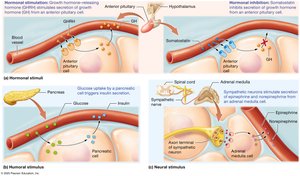
Hormonal stimuli: Endocrine cells respond to other hormones.
Humoral stimuli: Endocrine cells respond to ion/compound concentrations in blood/ECF.
Neural stimuli: Endocrine cells respond to nervous system signals.
Negative Feedback Regulation
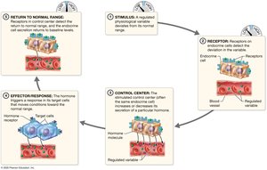
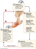
Hormone secretion is typically regulated by negative feedback loops:
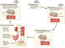
Stimulus: Variable deviates from normal range.
Receptor: Endocrine cell receptors detect deviation.
Control center: Endocrine cell adjusts hormone secretion.
Effector/response: Hormone triggers response to restore normal range.
Return to normal: Secretion returns to baseline.
Structure and Function of the Hypothalamus and Pituitary Gland
Anatomy of the Hypothalamus and Pituitary
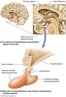
The hypothalamus connects to the pituitary gland via the infundibulum. The anterior pituitary (adenohypophysis) is a true gland, while the posterior pituitary (neurohypophysis) is nervous tissue that stores neurohormones.
Hormones of the Hypothalamus and Posterior Pituitary
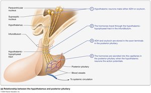
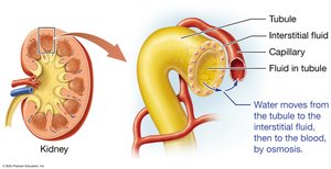
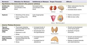
Antidiuretic hormone (ADH): Water retention; produced by hypothalamic neurons, stored in posterior pituitary, released in response to high solute concentration.
Oxytocin: Stimulates uterine contractions and milk ejection; works via positive feedback.
Functional Relationship of the Hypothalamus and Anterior Pituitary
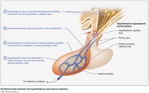
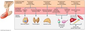
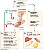
The hypothalamus produces releasing and inhibiting hormones that control the anterior pituitary. Tropic hormones from the anterior pituitary regulate other glands.
Anterior Pituitary Hormones
Thyroid-stimulating hormone (TSH): Stimulates thyroid gland.
Adrenocorticotropic hormone (ACTH): Stimulates adrenal cortex.
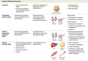
Prolactin: Stimulates mammary gland growth and milk production.
Luteinizing hormone (LH): Stimulates sex hormone production and ovulation.
Follicle-stimulating hormone (FSH): Stimulates gamete production.
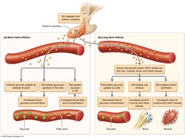
Growth hormone (GH): Promotes growth, protein synthesis, and metabolic effects.
Hormones of the Hypothalamus and Pituitary Gland (Table)
Hormone | Stimulus for Release | Inhibitor(s) of Release | Target Tissue(s) | Effects |
|---|---|---|---|---|
ADH | Increased solute concentration | Decreased solute concentration | Kidneys, brain | Water reabsorption, increased blood volume |
Oxytocin | Stretching of uterus, infant suckling | Lack of appropriate stimulus | Uterus, mammary gland | Uterine contractions, milk let-down reflex |
TSH | TRH from hypothalamus | Somatostatin | Thyroid gland | Growth and development, secretion of thyroid hormones |
ACTH | CRH from hypothalamus, stress | Increased level of corticosteroids | Adrenal cortex | Growth and development, secretion of steroid hormones |
Prolactin | Infant suckling, TRH from hypothalamus | Prolactin-inhibiting factor | Mammary gland | Development, milk production |
LH | GnRH from hypothalamus | Increased levels of testosterone/estrogens/progesterone | Male/female gonads | Development, production of hormones, ovulation |
FSH | GnRH from hypothalamus | Increased levels of inhibin | Male/female gonads | Production of gametes, maturation of ovarian follicles |
GH | GHRH from hypothalamus, protein ingestion, fasting | Somatostatin | Liver, adipose, bone, muscle | Growth, protein synthesis, breakdown of fat, gluconeogenesis |
Structure and Function of the Thyroid and Parathyroid Glands
Anatomy of the Thyroid and Parathyroid Glands
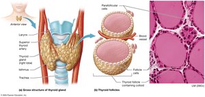
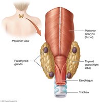
The thyroid gland is located in the anterior neck and consists of two lobes connected by the isthmus. It is composed of thyroid follicles, which produce thyroid hormones, and parafollicular cells, which produce calcitonin. The parathyroid glands are usually four small glands embedded in the posterior thyroid; chief cells produce parathyroid hormone (PTH).
Thyroid Hormones: Metabolic Regulators
Triiodothyronine (T3): Contains three iodine atoms; more physiologically active.
Thyroxine (T4): Contains four iodine atoms; converted to T3 in target cells.
Regulate basal metabolic rate, thermoregulation, growth, and development.
Synergize with the sympathetic nervous system.
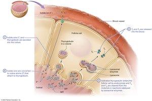
Thyroid Hormone Production
Iodide ions and thyroglobulin are secreted into the colloid.
Iodide is converted to iodine and attaches to thyroglobulin, forming MIT and DIT.
Iodinated thyroglobulin is endocytosed and cleaved to release T3 and T4.
Most T4 is bound to plasma proteins; only free T3 and T4 are biologically active.
Regulation of Thyroid Hormone Production
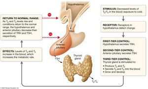
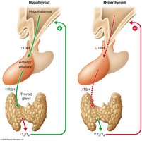
Regulated by a negative feedback loop involving TRH (hypothalamus), TSH (anterior pituitary), and thyroid hormones.
Thyroid Disorders
Hyperthyroidism: Overproduction of thyroid hormones; causes weight loss, heat intolerance, increased heart rate.
Hypothyroidism: Underproduction; causes weight gain, cold intolerance, slow heart rate.
Graves Disease: Autoimmune hyperthyroidism; may cause exophthalmos and goiter.
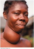
Goiter: Enlargement of the thyroid gland.
Congenital hypothyroidism: Inadequate thyroid function in infants; leads to developmental delays if untreated.
Negative Feedback Loops and Thyroid Function
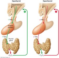
TSH secretion increases with hypothyroidism and decreases with hyperthyroidism.
In hypothyroidism, the thyroid cannot produce more T3/T4, so TRH and TSH remain elevated.
In hyperthyroidism, T3/T4 are elevated, so TRH and TSH fall to nearly zero.
Parathyroid Hormone and Calcitonin: Bone Homeostasis
Parathyroid hormone (PTH): Increases blood calcium by stimulating osteoclasts, increasing intestinal absorption, and increasing kidney reabsorption.
Calcitonin: Produced by thyroid parafollicular cells; inhibits osteoclasts, lowers blood calcium.
Hormones of the Thyroid and Parathyroid Glands (Table)
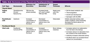
Cell Type | Hormone(s) | Stimulus for Secretion | Inhibitor(s) of Secretion | Target Tissue(s) | Effects |
|---|---|---|---|---|---|
Follicle cells | T3, T4 | TSH | Increased T3/T4 | Nearly every cell | Basal metabolic rate, thermoregulation, growth, development, SNS synergy |
Parafollicular cells | Calcitonin | Increased blood calcium | Decreased blood calcium | Osteoclasts | Inhibits osteoclasts, lowers blood calcium |
Chief cells | PTH | Decreased blood calcium | Increased blood calcium | Bone, kidneys, intestines | Increases blood calcium via bone resorption, kidney reabsorption, intestinal absorption |
 ----------------------------------------
