 Back
Chapter 17: Blood – Structure, Function, and Clinical Aspects
Study Guide - Smart Notes
 Tailored notes based on your materials, expanded with key definitions, examples, and context.
Blood: Internal Transport System
Overview
Blood is a life-sustaining fluid tissue that serves as the transport vehicle of the cardiovascular system. It is essential for delivering nutrients, removing wastes, and supporting homeostasis and immunity.
Transport: Delivers oxygen and nutrients, removes metabolic wastes, and transports hormones.
Regulation: Maintains temperature, pH, and fluid volume.
Protection: Prevents blood loss and infection via clotting and immune agents.
Composition of Blood
Main Components
Blood consists of plasma (the matrix) and formed elements (cells and cell fragments).
Plasma: Straw-colored fluid, about 90% water, contains dissolved solutes.
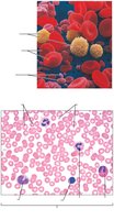
Formed Elements: Erythrocytes (RBCs), leukocytes (WBCs), and platelets.
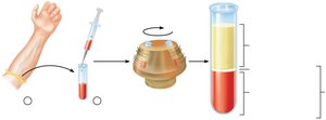
Physical Characteristics and Volume
Sticky, opaque fluid with metallic taste.
Color varies with oxygen content: scarlet red (high O2), dark red (low O2).
pH: 7.35–7.45; makes up ~8% of body weight.
Average volume: Males 5–6 L, Females 4–5 L.
Blood Plasma
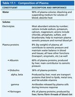
Plasma is the liquid portion of blood, containing water, proteins, nutrients, gases, hormones, and wastes.
Albumin: Most abundant plasma protein; maintains osmotic pressure and acts as a carrier.
Globulins: Transport proteins and antibodies.
Fibrinogen: Essential for blood clotting.
Constituent | Description and Importance |
|---|---|
Water | 90% of plasma volume; dissolving and suspending medium for solutes of blood; absorbs heat |
Electrolytes | Most abundant solutes by number; help maintain osmotic pressure, pH, and membrane potential |
Plasma proteins | Albumin (60%), globulins (36%), fibrinogen (4%) |
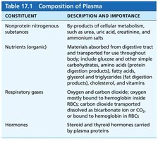
Constituent | Description and Importance |
|---|---|
Nonprotein nitrogenous substances | By-products of cellular metabolism |
Nutrients (organic) | Materials absorbed from digestive tract |
Respiratory gases | Oxygen and carbon dioxide |
Hormones | Steroid and thyroid hormones carried by plasma proteins |
Formed Elements
Types and Characteristics
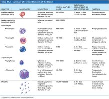
Erythrocytes (RBCs): No nuclei or organelles; dedicated to gas transport.
Leukocytes (WBCs): Complete cells; defense against disease.
Platelets: Cell fragments; essential for clotting.
Erythrocytes (Red Blood Cells)
Structural Characteristics
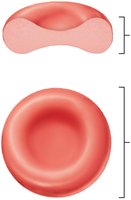
Erythrocytes are small, biconcave, anucleate cells optimized for gas transport.
Biconcave shape increases surface area for gas exchange.
Filled with hemoglobin (Hb), the oxygen-carrying protein.
No mitochondria; ATP production is anaerobic.
Function of Erythrocytes
Dedicated to respiratory gas transport.
Hemoglobin binds reversibly with oxygen.
Each Hb molecule can transport four O2 molecules.
Structure of Hemoglobin
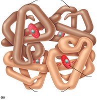
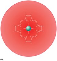
Hemoglobin consists of four polypeptide chains (two alpha, two beta) and four heme groups, each with an iron atom that binds O2. 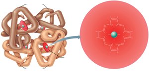
Production of Erythrocytes (Erythropoiesis)
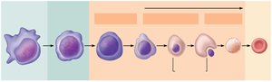
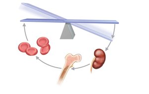
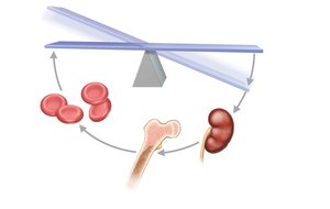
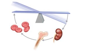
Erythropoiesis is the process of RBC formation, occurring in red bone marrow.
Stem cell (hemocytoblast) differentiates into committed cells.
Stages: proerythroblast → basophilic erythroblast → polychromatic erythroblast → orthochromatic erythroblast → reticulocyte → mature erythrocyte.
Regulation of Erythropoiesis
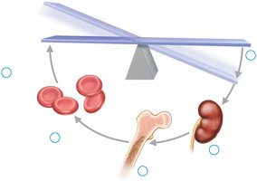
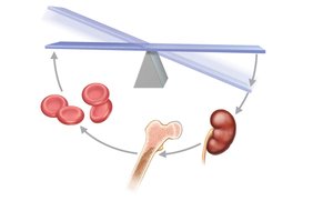
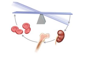
Controlled by erythropoietin (EPO), a hormone released by kidneys in response to hypoxia.
Balance between RBC production and destruction is crucial.
Dietary requirements: amino acids, iron, vitamin B12, folic acid.
Fate and Destruction of Erythrocytes
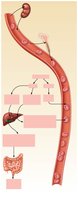
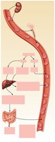
RBCs have a lifespan of 100–120 days. Old RBCs are removed by macrophages in the spleen, liver, and bone marrow.
Hemoglobin is broken down: iron is stored/reused, heme is converted to bilirubin, globin is metabolized to amino acids.
Erythrocyte Disorders
Anemia
Anemia is a condition of abnormally low O2-carrying capacity.
Blood loss: Hemorrhagic anemia (acute or chronic).
Not enough RBCs produced: Iron-deficiency, pernicious, renal, aplastic anemia.
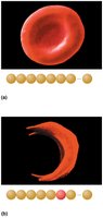
Too many RBCs destroyed: Hemolytic anemias, thalassemias, sickle-cell anemia.
Polycythemia
Polycythemia is an abnormal excess of RBCs, increasing blood viscosity.
Polycythemia vera: bone marrow cancer.
Secondary polycythemia: response to low O2 or increased EPO.
Blood doping: artificial increase in RBCs for athletic performance.
Leukocytes (White Blood Cells)
General Structure and Function
Leukocytes are complete cells that defend against disease.
Can leave capillaries (diapedesis) and move through tissues.
Leukocytosis: elevated WBC count, normal response to infection.
Types of Leukocytes
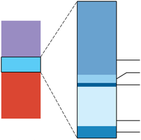
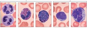
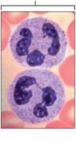
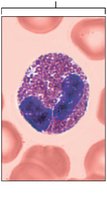
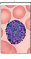
Granulocytes: Neutrophils, eosinophils, basophils (contain granules).
Agranulocytes: Lymphocytes, monocytes (lack granules).
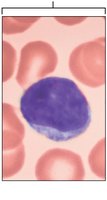
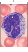
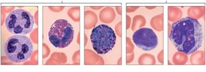
Leukocyte Formation (Leukopoiesis)
Leukopoiesis is stimulated by interleukins and colony-stimulating factors.
All leukocytes originate from hemocytoblast stem cells.
Lymphoid stem cells produce lymphocytes; myeloid stem cells produce other WBCs.
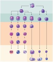
Platelets
Structure and Function
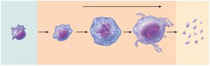
Platelets are fragments of megakaryocytes, essential for clotting.
Contain chemicals for clotting: serotonin, calcium, enzymes, ADP.
Form temporary platelet plugs to seal vessel breaks.
Cell Type | Description | Function |
|---|---|---|
Erythrocyte | Biconcave, anucleate | Transport O2 and CO2 |
Leukocyte | Complete cell | Defense against disease |
Platelet | Cell fragment | Clotting |
Hemostasis
Steps in Hemostasis
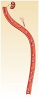
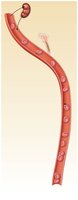
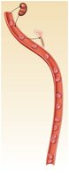
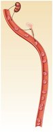
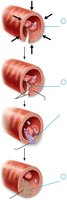
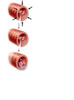
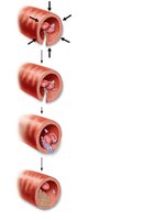
Hemostasis is the process of stopping bleeding, involving three steps:
Vascular spasm: Vasoconstriction of injured vessel.
Platelet plug formation: Platelets adhere to exposed collagen and release chemicals.
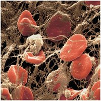
Coagulation: Reinforcement of plug with fibrin threads.
Coagulation Pathways
Intrinsic pathway: triggered by factors within blood.
Extrinsic pathway: triggered by tissue factor outside blood.
Both pathways lead to formation of prothrombin activator, conversion of prothrombin to thrombin, and formation of fibrin mesh.
Clot Retraction and Fibrinolysis
Clot retraction: platelets contract, pulling vessel edges together.
Fibrinolysis: plasmin digests fibrin, dissolving the clot.
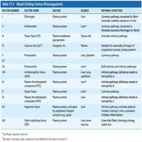
Factor Number | Factor Name | Source | Function |
|---|---|---|---|
I | Fibrinogen | Liver | Converted to fibrin |
II | Prothrombin | Liver | Converted to thrombin |
IV | Calcium ions | Diet | Essential for clotting |
Disorders of Hemostasis
Thromboembolic Disorders
Thrombus: Clot in unbroken vessel.
Embolus: Clot that travels in bloodstream.
Risk factors: atherosclerosis, inflammation, immobility.
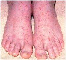
Bleeding Disorders
Impaired liver function: Inability to synthesize clotting factors.
Hemophilia: Hereditary deficiency of clotting factors.
DIC: Widespread clotting and bleeding.
Blood Transfusions
ABO Blood Groups
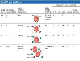
Blood groups are determined by antigens (agglutinogens) on RBC surfaces.
Type A: A antigen
Type B: B antigen
Type AB: A and B antigens
Type O: No antigens
Blood Group | RBC Antigens | Plasma Antibodies | Blood That Can Be Received |
|---|---|---|---|
A | A | Anti-B | A, O |
B | B | Anti-A | B, O |
AB | A, B | None | A, B, AB, O |
O | None | Anti-A, Anti-B | O |
Blood Typing
Blood typing involves mixing blood with antibodies to detect agglutination. 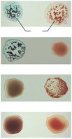
Diagnostic Blood Tests
Clinical Applications
Hematocrit, blood glucose, and WBC counts are used to diagnose anemia, diabetes, and infection.
Complete blood count (CBC) assesses overall blood health.
Developmental Aspects of Blood
Fetal and Aging Blood
Fetal blood cells form in yolk sac, liver, and spleen; red bone marrow becomes primary site by seventh month.
Hemoglobin F has higher O2 affinity than adult hemoglobin.
Blood diseases of aging include chronic leukemias, anemias, and clotting disorders.
