 Back
Chapter 17: The Circulatory System – Blood
Study Guide - Smart Notes
 Tailored notes based on your materials, expanded with key definitions, examples, and context.
Functions and Properties of Blood
Overview of Blood Functions
Blood is a vital connective tissue that performs numerous essential functions in the human body, including respiration, nutrient transport, waste elimination, thermoregulation, immune defense, and maintenance of water and pH balance. The average adult has 1–2 gallons of blood, which consists of plasma (the clear extracellular fluid) and formed elements (cells and platelets).
Viscosity: Refers to the resistance of blood to flow. High viscosity increases blood pressure, while low viscosity can cause edema.
Osmolarity: The total molarity of dissolved particles in blood. Imbalances can lead to hypertension or tissue swelling.
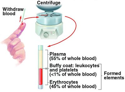
Composition of Blood
Plasma: Makes up about 55% of blood volume and contains proteins, enzymes, nutrients, wastes, hormones, and gases.
Formed Elements: Comprise about 45% of blood and include erythrocytes (RBCs), leukocytes (WBCs), and platelets.
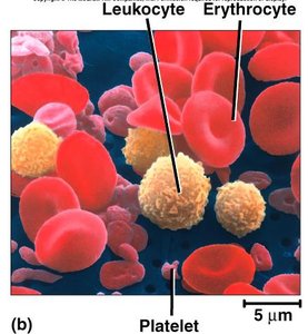
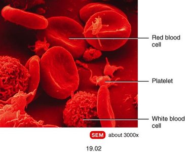
Hematocrit
Hematocrit is the percentage of total blood volume occupied by red blood cells. It is a key indicator of oxygen-carrying capacity.
Plasma and Plasma Proteins
Major Plasma Proteins
Albumins: Most abundant; contribute to viscosity, osmolarity, and blood pressure.
Globulins: Provide immune defenses (antibodies).
Fibrinogen: Precursor to fibrin, essential for blood clotting.
All plasma proteins except globulins are synthesized by the liver.
Nonprotein Components
Nitrogenous compounds (amino acids, urea)
Nutrients (glucose, vitamins, fats, minerals)
Gases (O2, CO2)
Electrolytes (mainly sodium ions)
Blood Cell Production (Hemopoiesis)
Sites and Regulation of Hemopoiesis
Blood cells are produced in hemopoietic tissues. In adults, red bone marrow is the primary site, while lymphoid tissues (thymus, tonsils, lymph nodes, spleen, Peyer's patches) are involved in lymphocyte production. In embryos, the yolk sac, liver, spleen, and thymus are involved.
Erythropoietin (EPO): Stimulates RBC production.
Thrombopoietin: Stimulates platelet production.
Colony-Stimulating Factors (CSFs): Stimulate WBC production.
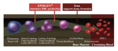
Erythropoiesis (Red Blood Cell Production)
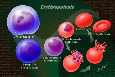
Erythropoiesis is the process of RBC formation, beginning with a proerythroblast, which responds to EPO. The cell undergoes several stages, losing its nucleus and becoming a reticulocyte before maturing into an erythrocyte. This process takes 3–5 days.
Regulation of Erythrocyte Homeostasis
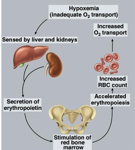
Negative feedback mechanisms regulate RBC production. Hypoxemia (low blood oxygen) stimulates the kidneys to release EPO, which increases RBC production in the bone marrow.
Nutritional Requirements for Erythropoiesis
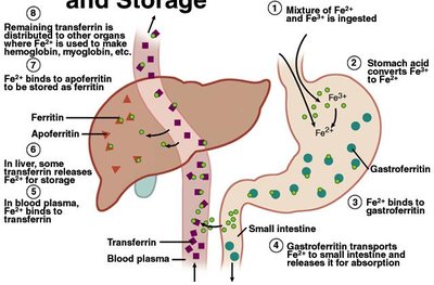
Iron: Essential for hemoglobin synthesis; absorbed as Fe2+ and transported by transferrin.
Vitamins B12 and Folic Acid: Required for DNA synthesis and rapid cell division.
Vitamin C and Copper: Cofactors for enzymes involved in RBC synthesis.
Formed Elements of Blood
Types of Formed Elements
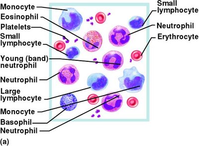
Erythrocytes (RBCs): Transport oxygen and carbon dioxide.
Leukocytes (WBCs): Immune defense; include granulocytes (neutrophils, eosinophils, basophils) and agranulocytes (lymphocytes, monocytes).
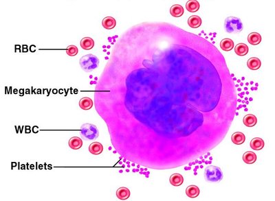
Platelets (Thrombocytes): Involved in clotting and vessel repair.
Erythrocytes and Hemoglobin
Structure and Function
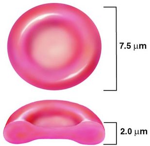
Erythrocytes are biconcave, disc-shaped cells specialized for gas transport. They lack organelles, maximizing space for hemoglobin, which binds and transports oxygen. The enzyme carbonic anhydrase in RBCs helps regulate pH by converting CO2 to carbonic acid.
Hemoglobin Structure
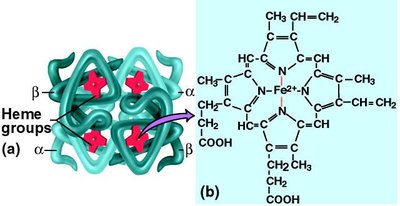
Hemoglobin consists of four protein chains (globins), each with a heme group that binds oxygen. Adult hemoglobin has two alpha and two beta chains, while fetal hemoglobin has gamma chains instead of beta.
RBC Count and Hemoglobin Concentration
Indicators of blood's oxygen-carrying capacity.
Lower in women due to menstrual losses and hormonal differences.
Erythrocyte Life Cycle and Disposal
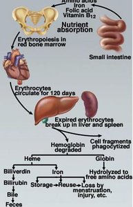
RBCs circulate for about 120 days before being broken down in the spleen and liver. Hemoglobin is degraded, with iron recycled and heme converted to bilirubin, which is excreted in bile.
Erythrocyte Disorders
Polycythemia
Polycythemia is an abnormal increase in RBC count, which can result from increased production, hypoxia, dehydration, or blood doping. It increases blood viscosity and risk of embolism, stroke, or heart failure.
Anemia
Anemia is a deficiency of RBCs or hemoglobin, leading to reduced oxygen delivery to tissues. Causes include inadequate erythropoiesis, hemorrhage, or hemolysis. Symptoms include fatigue, pallor, and shortness of breath.
Iron-deficiency anemia: Most common, especially in women.
Vitamin-deficiency anemia: Due to lack of B12, folate, or other vitamins.
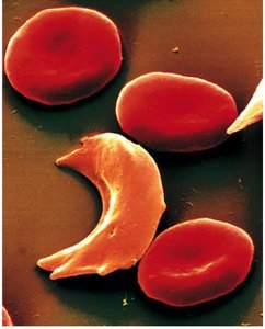
Sickle-cell disease: Hereditary disorder causing abnormal hemoglobin and sickle-shaped RBCs.
Blood Types
ABO and Rh Blood Groups
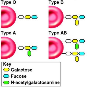
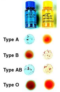
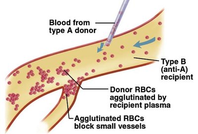
Blood types are determined by the presence or absence of specific antigens (agglutinogens) on RBCs. The ABO system is based on A and B antigens, while the Rh system is based on the D antigen.
Type A: A antigens, anti-B antibodies
Type B: B antigens, anti-A antibodies
Type AB: Both A and B antigens, no anti-A/B antibodies (universal recipient)
Type O: No A/B antigens, both anti-A and anti-B antibodies (universal donor)
Rh Factor and Hemolytic Disease of the Newborn
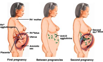
The Rh factor is another important antigen. Rh-negative individuals can develop antibodies if exposed to Rh-positive blood, which can cause hemolytic disease in newborns if the mother is Rh-negative and the fetus is Rh-positive.
Leukocytes (White Blood Cells)
Types and Functions
Granulocytes:
Neutrophils: Phagocytize bacteria, release antimicrobial chemicals.
Eosinophils: Combat parasites and allergens.
Basophils: Release histamine and heparin.
Agranulocytes:
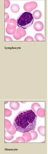
Lymphocytes: Destroy infected/cancerous cells, coordinate immune response, secrete antibodies.
Monocytes: Differentiate into macrophages, phagocytize pathogens and debris.
Leukocyte Disorders
Leukopenia: Low WBC count, increased infection risk.
Leukocytosis: High WBC count, often due to infection or inflammation.
Leukemia: Cancer of blood-forming tissues, leading to abnormal WBC proliferation.
Platelets and Hemostasis
Platelet Structure and Function
Platelets are small fragments of megakaryocyte cytoplasm. They play a crucial role in hemostasis by forming plugs, secreting clotting factors, and attracting WBCs to sites of inflammation.
Hemostasis: The Control of Bleeding
Hemostasis is the process of stopping bleeding and involves three main steps:
Vascular Spasm: Constriction of the damaged vessel to reduce blood flow.
Platelet Plug Formation: Platelets adhere to exposed collagen and aggregate to form a temporary plug.
Coagulation (Clotting): Fibrinogen is converted to fibrin, forming a stable clot.
Coagulation Pathways
Clotting involves a cascade of reactions with two pathways:
Extrinsic Pathway: Initiated by tissue thromboplastin.
Intrinsic Pathway: Initiated by factors within the blood.
Both pathways converge to activate factor X, leading to the conversion of prothrombin to thrombin, and fibrinogen to fibrin.
Clot Retraction and Dissolution
Clot Retraction: Platelets contract to condense the clot.
Fibrinolysis: Plasmin dissolves the fibrin meshwork, breaking down the clot.
Prevention of Inappropriate Coagulation
Platelet repulsion by prostacyclin-coated endothelium
Thrombin dilution by rapid blood flow
Natural anticoagulants (antithrombin, heparin)
Coagulation Disorders
Hemophilia: Genetic deficiency of clotting factors, leading to excessive bleeding.
Thrombosis: Abnormal clotting in unbroken vessels, which can lead to embolism and infarction (tissue death).
Example: Hemophilia A is due to a deficiency of factor VIII and is the most common form of hemophilia.
Additional info: Vitamin K is essential for the synthesis of several clotting factors. Deficiency can lead to bleeding disorders.
