 Back
BackChapter 18: The Circulatory System – Blood (Anatomy & Physiology Study Notes)
Study Guide - Smart Notes
 Tailored notes based on your materials, expanded with key definitions, examples, and context.
The Circulatory System: Blood
General Aspects of Blood
The circulatory system is essential for transporting substances throughout the body, protecting against disease, and regulating internal conditions. Blood, a liquid connective tissue, is composed of plasma and formed elements, each with distinct functions and properties.
Circulatory System: Includes the heart, blood vessels, and blood.
Cardiovascular System: Refers to the heart and blood vessels only.
Hematology: The study of blood.
Functions:
Transport: O2, CO2, nutrients, wastes, hormones, stem cells.
Protection: Inflammation, infection control, toxin neutralization, clotting.
Regulation: Fluid balance, pH stabilization, temperature control.
Components and Properties of Blood
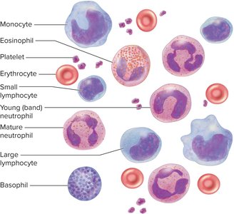
Blood consists of plasma (the matrix) and formed elements (cells and cell fragments). The formed elements include erythrocytes (RBCs), platelets, and leukocytes (WBCs). Granulocytes (neutrophils, eosinophils, basophils) and agranulocytes (lymphocytes, monocytes) are types of WBCs.
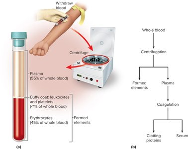
Blood Fractionation: Separation of blood into components by centrifugation and coagulation.
Hematocrit: Percentage of blood volume composed of RBCs (typically 37–52%).
Serum: Plasma without clotting proteins.
Blood Plasma
Plasma is a complex mixture of water, proteins, nutrients, electrolytes, wastes, hormones, and gases. Plasma proteins are the most abundant solutes by weight.
Albumins: Smallest, most abundant; transport solutes, buffer pH, contribute to viscosity and osmotic pressure.
Globulins: Alpha, beta, gamma; roles in transport, clotting, immunity.
Fibrinogen: Soluble precursor to fibrin, forms blood clot framework.
Nitrogenous Wastes: Urea is the most abundant, removed by kidneys.
Nutrients: Glucose, amino acids, fats, cholesterol, vitamins, minerals.
Electrolytes: Sodium ions (Na+) are predominant.
Blood Viscosity and Osmolarity
Viscosity and osmolarity are critical for circulatory function. Viscosity refers to resistance to flow, while osmolarity is the concentration of solute particles.
Viscosity: Whole blood is 4.5–5.5 times as viscous as water; plasma is 2 times as viscous.
Osmolarity: Regulated by sodium ions, proteins, RBCs; affects blood pressure and tissue fluid balance.
Colloid Osmotic Pressure (COP): Protein contribution to osmotic pressure, important for water balance.
Starvation and Plasma Protein Deficiency
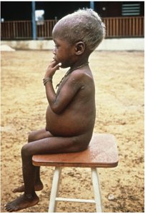
Hypoproteinemia, a deficiency of plasma proteins, can result from starvation, liver/kidney disease, or burns. It leads to fluid loss from blood to tissues, causing swelling and ascites. Kwashiorkor is a severe protein deficiency seen in malnourished children.
How Blood Is Produced
Blood components are continually replaced through hematopoiesis. Hematopoietic tissues produce blood cells, with myeloid hematopoiesis occurring in red bone marrow and lymphoid hematopoiesis in lymphoid organs.
Hematopoietic Stem Cells (HSCs): Multipotent cells in bone marrow, give rise to all formed elements.
Colony-Forming Units (CFUs): Specialized progenitors for specific blood cell types.
Plasma Replacement: Water, nutrients, electrolytes absorbed from digestive tract; proteins mainly from liver.
Erythrocytes (Red Blood Cells)
Structure and Function
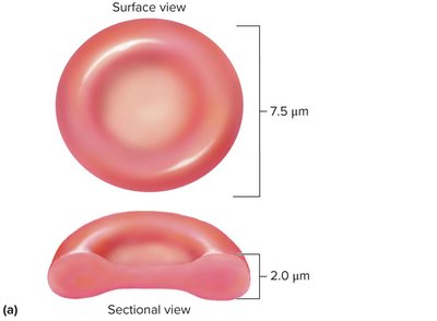
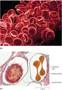
Erythrocytes are discoid cells with a biconcave shape, specialized for gas transport. They lack mitochondria, nucleus, and DNA, relying on anaerobic fermentation for ATP production.
Diameter: 7.5 μm; thickness: 2.0 μm at rim.
Hemoglobin: Constitutes 33% of cytoplasm; 280 million molecules per RBC.
Carbonic Anhydrase: Enzyme for CO2 transport and pH balance.
Glycolipids: Determine blood type.
Cytoskeletal Proteins: Spectrin and actin provide resilience and durability.
Hemoglobin
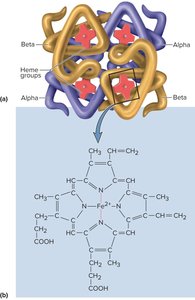
Hemoglobin is a protein with four globin chains (two alpha, two beta; fetal hemoglobin has gamma chains). Each chain is associated with a heme group that binds iron and carries oxygen.
Each hemoglobin: Can transport up to four O2 molecules.
CO2 Transport: About 5% of CO2 binds to globin.
Quantities of Erythrocytes and Hemoglobin
Clinical measurements assess oxygen-carrying capacity:
Hematocrit: Men: 42–52%; Women: 37–48%.
Hemoglobin Concentration: Men: 13–18 g/dL; Women: 12–16 g/dL.
RBC Count: Men: 4.6–6.2 million/μL; Women: 4.2–5.4 million/μL.
Erythrocyte Life History
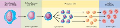
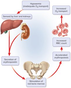
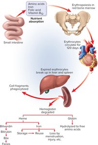
Erythropoiesis is the process of RBC production, regulated by erythropoietin (EPO) from the kidneys. RBCs have a lifespan of about 120 days.
Stages: HSC → CFU → Erythroblast → Reticulocyte → Mature RBC.
Negative Feedback: Hypoxemia stimulates EPO, increasing RBC production.
Iron Metabolism
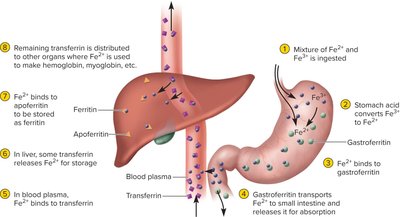
Iron is essential for hemoglobin synthesis and is absorbed, transported, and stored through specialized proteins.
Dietary Iron: Ferric (Fe3+) and ferrous (Fe2+) forms.
Gastroferritin: Binds Fe2+ for absorption.
Transferrin: Transports iron in blood.
Ferritin: Storage form in liver.
Erythrocyte Death and Disposal
RBCs are removed by macrophages in the spleen and liver. Hemoglobin is degraded, and its components are recycled or excreted.
Globin: Hydrolyzed to amino acids.
Heme: Converted to biliverdin, then bilirubin; excreted in urine and bile.
Erythrocyte Disorders
Disorders include polycythemia (excess RBCs), anemia (deficiency of RBCs or hemoglobin), and sickle-cell disease (hereditary hemoglobin defect).
Polycythemia: Can result from cancer, dehydration, high altitude, or physical conditioning.
Anemia: Causes include hemorrhage, hemolysis, inadequate erythropoiesis, iron deficiency, and autoimmune disorders.
Sickle-cell Disease: HbS variant causes RBCs to become rigid and block vessels; heterozygotes are malaria-resistant.
Blood Types
ABO Blood Group
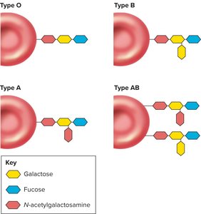
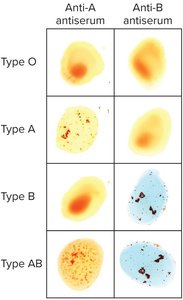
Blood types are determined by antigens (agglutinogens) on RBCs and antibodies (agglutinins) in plasma. The ABO group includes types A, B, AB, and O.
Type A: Antigen A present.
Type B: Antigen B present.
Type AB: Both antigens present.
Type O: Neither antigen present.
Agglutination and Transfusion Compatibility
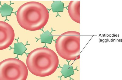
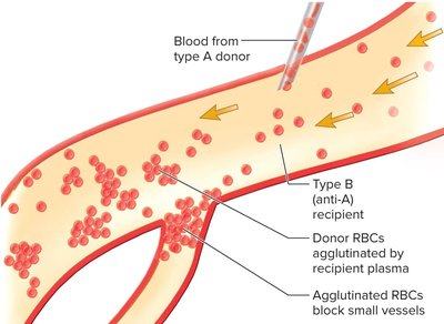
Antibodies react against foreign antigens, causing agglutination. Transfusion reactions occur if donor RBCs are agglutinated by recipient plasma.
Universal Donor and Recipient
Type AB: Universal recipient (no anti-A or anti-B antibodies).
Type O: Universal donor (no antigens on RBCs).
Rh Blood Group
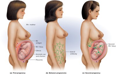
The Rh group includes antigens C, D, and E. Rh-positive (Rh+) individuals have antigen D; Rh-negative (Rh−) lack it. Anti-D antibodies form only after exposure to Rh+ blood.
Maternal-Fetal Mismatches
Hemolytic disease of the newborn (HDN) can occur when an Rh− mother has an Rh+ fetus. Anti-D antibodies can cross the placenta and cause fetal anemia. Prevention involves Rh immune globulin (RhoGAM).
Leukocytes (White Blood Cells)
Form and Function
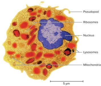
Leukocytes protect against infection and disease. They are the least abundant formed element and migrate into tissues after a few hours in the bloodstream.
Granulocytes: Neutrophils, eosinophils, basophils (contain specific granules).
Agranulocytes: Lymphocytes, monocytes (lack specific granules).
Types of Leukocytes
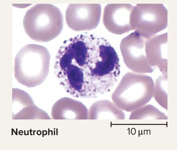
Neutrophils: 60–70%; antibacterial, phagocytize bacteria, release antimicrobial chemicals.
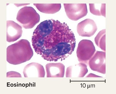
Eosinophils: 2–4%; phagocytize antigen-antibody complexes, release enzymes against parasites.
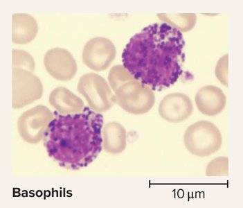
Basophils: <0.5%; secrete histamine (vasodilator) and heparin (anticoagulant).
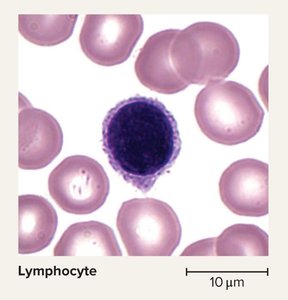
Lymphocytes: 25–33%; destroy cancer/foreign/virally infected cells, coordinate immune response, secrete antibodies.
Monocytes: 3–8%; transform into macrophages, phagocytize pathogens/debris, act as antigen-presenting cells.
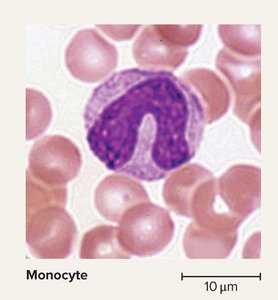
Leukocyte Life History
Leukopoiesis is the production of WBCs from hematopoietic stem cells. Myeloblasts form granulocytes, monoblasts form monocytes, and lymphoblasts form lymphocytes.
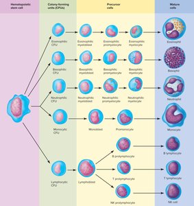
Leukocyte Disorders
Leukopenia: Low WBC count; increased infection risk.
Leukocytosis: High WBC count; often due to infection, allergy, disease.
Leukemia: Cancer of hematopoietic tissue; uncontrolled WBC production, impaired clotting, infections.
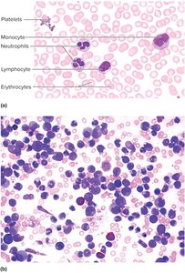
Platelets and Control of Bleeding
Platelet Form and Function
Platelets are small fragments of megakaryocytes, essential for hemostasis (cessation of bleeding). They lack a nucleus and contain granules with secretions.
Functions: Vasoconstriction, platelet plug formation, clotting factor secretion, clot dissolution, inflammation, growth factor secretion.
Thrombocytopenia: Platelet deficiency.
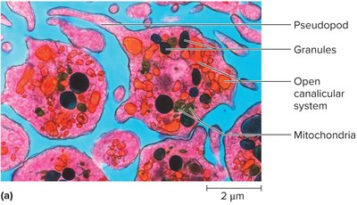
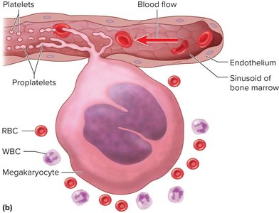
Platelet Production
Thrombopoiesis is triggered by thrombopoietin, leading to megakaryocyte formation. Platelets are fragments released into the bloodstream.
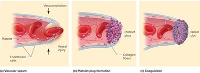
Hemostasis
Hemostasis involves three mechanisms: vascular spasm, platelet plug formation, and coagulation.
Vascular Spasm: Immediate vessel constriction after injury.
Platelet Plug Formation: Platelets adhere to exposed collagen, aggregate, and release factors.
Coagulation: Conversion of fibrinogen to fibrin, forming a clot.
Coagulation Pathways
Coagulation occurs via extrinsic (tissue thromboplastin) and intrinsic (platelet factor 12) pathways, both requiring calcium and leading to a common pathway.
Fate of Blood Clots
After clot formation, tissue repair begins. Platelet-derived growth factor stimulates healing, and fibrinolysis dissolves the clot.
Prevention of Inappropriate Clotting
Platelet Repulsion: Platelets do not adhere to prostacyclin-coated endothelium.
Dilution: Thrombin is diluted by blood flow.
Anticoagulants: Antithrombin and heparin inhibit clotting.
Clotting Disorders
Hemophilia: Hereditary deficiency of clotting factors; most common types are sex-linked.
Thrombosis: Abnormal clot formation in unbroken vessels; risk of embolism and infarction.
Clinical Management: Vitamin K antagonists, streptokinase, tissue plasminogen activator, hementin.
