 Back
Chapter 18: The Endocrine System – Fundamentals of Anatomy & Physiology
Study Guide - Smart Notes
 Tailored notes based on your materials, expanded with key definitions, examples, and context.
The Endocrine System
Introduction to the Endocrine System
The endocrine system is a major regulatory system of the body, responsible for producing and releasing hormones that control and coordinate various physiological processes. Endocrine cells and tissues produce about 30 different hormones, which act as chemical messengers to influence the function of target organs and maintain homeostasis.
Endocrine cells: Specialized cells that secrete hormones directly into the bloodstream.
Hormones: Chemical messengers that regulate activities such as growth, metabolism, and reproduction.
Homeostasis: The maintenance of stable internal conditions.
Mechanisms of Intercellular Communication
Intercellular communication is essential for coordinating body functions. The endocrine and nervous systems use different mechanisms to transmit signals between cells.
Direct communication: Exchange of ions and molecules between adjacent cells via gap junctions; rare and highly specialized.
Paracrine communication: Chemical signals transfer information between cells within a single tissue.
Autocrine communication: Cells respond to signals they themselves produce (e.g., prostaglandins).
Endocrine communication: Hormones are released into the bloodstream and affect distant target cells.
Synaptic communication: Neurons release neurotransmitters at synapses for rapid, targeted responses.
Hormones and Their Actions
Classes of Hormones
Hormones are classified based on their chemical structure, which determines their mechanism of action and transport.
Amino acid derivatives: Small molecules derived from amino acids (e.g., thyroid hormones, catecholamines, serotonin, melatonin).
Peptide hormones: Chains of amino acids, including glycoproteins and small polypeptides (e.g., TSH, LH, FSH, insulin, ADH, OXT).
Lipid derivatives: Includes eicosanoids (from arachidonic acid) and steroid hormones (from cholesterol, e.g., androgens, estrogens, corticosteroids, calcitriol).
Hormone Transport and Inactivation
Hormones may circulate freely or be bound to carrier proteins. Free hormones are quickly inactivated, while bound hormones remain functional longer.
Free hormones: Functional for less than an hour; inactivated by binding, breakdown, or enzymatic degradation.
Bound hormones: Attached to transport proteins; provide a reserve in the bloodstream.
Mechanisms of Hormone Action
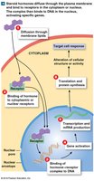
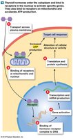
Hormones exert their effects by binding to specific receptors on or within target cells, altering genetic activity, protein synthesis, or membrane permeability.
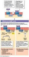
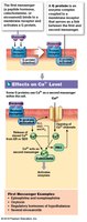
Extracellular receptors: For peptide hormones and catecholamines; activate second messenger systems (e.g., cAMP, Ca2+).
Intracellular receptors: For steroid and thyroid hormones; directly affect gene expression and protein synthesis.
Regulation of Hormone Secretion
Hormone secretion is primarily controlled by negative feedback mechanisms, triggered by humoral, hormonal, or neural stimuli.
Humoral stimuli: Changes in extracellular fluid.
Hormonal stimuli: Arrival or removal of another hormone.
Neural stimuli: Neurotransmitter signals.
The Pituitary Gland
Anatomy and Function
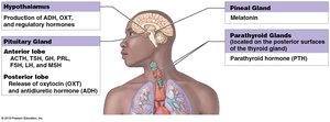
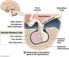
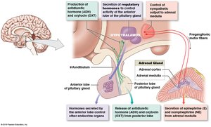
The pituitary gland (hypophysis) is located within the sella turcica, connected to the hypothalamus by the infundibulum. It releases nine peptide hormones that regulate various bodily functions.
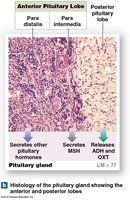
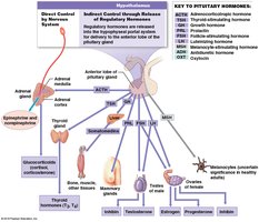
Anterior lobe (adenohypophysis): Produces hormones that "turn on" other endocrine glands or support organ functions.
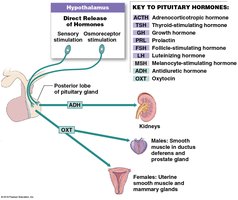
Posterior lobe (neurohypophysis): Releases ADH and OXT synthesized by the hypothalamus.
Hypothalamic Control
The hypothalamus regulates pituitary function through three mechanisms: secretion of regulatory hormones, production of ADH and OXT, and direct neural control of the adrenal medulla.
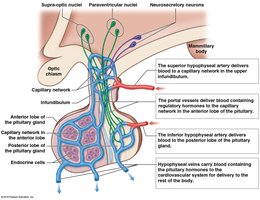
Hypophyseal Portal System
Portal vessels link two capillary networks, ensuring regulatory hormones reach the anterior pituitary before entering general circulation.
Feedback Control of Endocrine Secretion
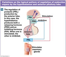
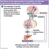
Negative feedback regulates hormone secretion, involving releasing and inhibiting hormones from the hypothalamus.
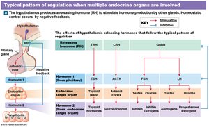
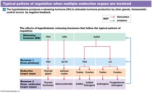
Hormones of the Anterior Pituitary
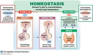
TSH: Stimulates thyroid gland.
ACTH: Stimulates adrenal cortex.
PRL: Stimulates mammary glands.
GH: Stimulates growth and metabolism.
FSH & LH: Regulate gonadal function.
MSH: Stimulates melanin production (pars intermedia).
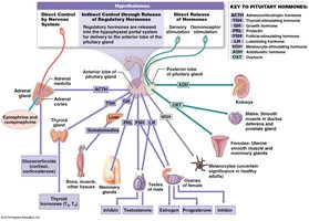
Hormones of the Posterior Pituitary
ADH: Regulates water balance.
OXT: Stimulates uterine contraction and milk ejection.
The Thyroid Gland
Anatomy and Function
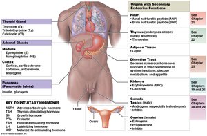
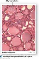
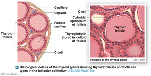
The thyroid gland is located inferior to the thyroid cartilage and consists of two lobes connected by an isthmus. It contains thyroid follicles and parafollicular (C) cells.
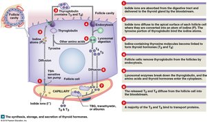
Thyroid follicles: Produce thyroglobulin, the precursor to thyroid hormones.
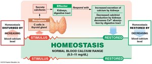
C cells: Produce calcitonin, which regulates calcium levels.
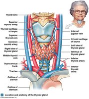
Thyroid Hormones
Thyroxine (T4): Contains four iodine atoms.
Triiodothyronine (T3): Contains three iodine atoms.
Thyroid-binding globulins (TBGs): Transport thyroid hormones in the blood.
Regulation and Effects of Thyroid Hormones
Essential for normal development of skeletal, muscular, and nervous systems in children.
Increase metabolic rate, energy consumption, and heat production (calorigenic effect).
Regulate heart rate, respiratory sensitivity, red blood cell formation, and bone mineral turnover.
Calcitonin
Produced by C cells.
Reduces blood calcium levels by stimulating excretion and inhibiting absorption.
Parathyroid Glands
Anatomy and Function
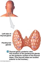
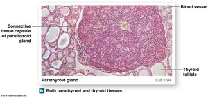
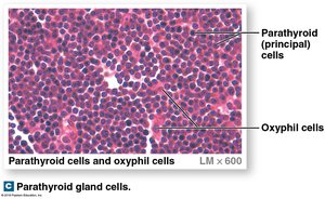
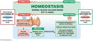
The parathyroid glands are four small glands embedded in the posterior surface of the thyroid gland. They secrete parathyroid hormone (PTH) in response to low blood calcium levels.
PTH: Stimulates osteoclasts, enhances calcium reabsorption by kidneys, and promotes calcitriol synthesis.
Adrenal Glands
Anatomy and Function
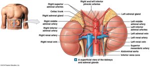
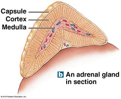
The adrenal glands are located on the superior border of each kidney and consist of an outer cortex and inner medulla.
Adrenal cortex: Produces corticosteroids (mineralocorticoids, glucocorticoids, and androgens).
Adrenal medulla: Produces catecholamines (epinephrine and norepinephrine).
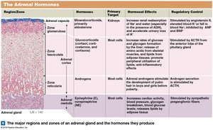
Zones of the Adrenal Cortex
Zona glomerulosa: Produces mineralocorticoids (aldosterone).
Zona fasciculata: Produces glucocorticoids (cortisol, corticosterone, cortisone).
Zona reticularis: Produces androgens.
Adrenal Medulla
Produces epinephrine (E) and norepinephrine (NE).
Mobilizes energy reserves, increases heart rate, and prepares the body for "fight or flight" responses.
Pineal Gland
Anatomy and Function
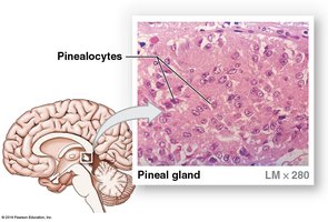
The pineal gland is located in the posterior portion of the roof of the third ventricle. Pinealocytes synthesize melatonin, which regulates circadian rhythms, inhibits reproductive functions, and protects against free radicals.
Pancreas
Anatomy and Function
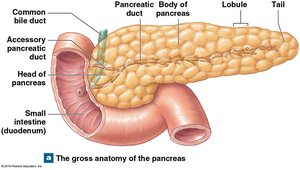
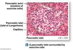
The pancreas is a large gland located between the stomach and small intestine. It contains both exocrine and endocrine cells.
Exocrine pancreas: Secretes digestive enzymes.
Endocrine pancreas: Contains pancreatic islets (alpha, beta, delta, and PP cells).
Hormones of the Pancreas
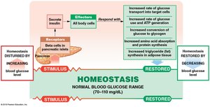
Insulin: Produced by beta cells; lowers blood glucose by promoting uptake and storage.
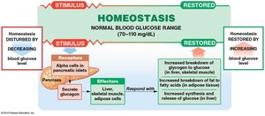
Glucagon: Produced by alpha cells; raises blood glucose by promoting glycogen breakdown and gluconeogenesis.
Diabetes Mellitus
Type 1 diabetes: Inadequate insulin production; requires insulin therapy.
Type 2 diabetes: Insulin resistance; often associated with obesity.
Complications: Kidney degeneration, retinal damage, heart attacks, neuropathies, tissue damage.
Secondary Endocrine Functions
Other Organs with Endocrine Functions
Intestines: Release hormones for digestive coordination.
Kidneys: Produce calcitriol, erythropoietin (EPO), and renin.
Heart: Produces natriuretic peptides (ANP, BNP).
Thymus: Produces thymosin for lymphocyte development.
Gonads: Testes produce androgens and inhibin; ovaries produce estrogens, progesterone, and inhibin.
Adipose tissue: Produces leptin for appetite regulation.
Hormone Interactions and General Adaptation Syndrome
Types of Hormone Interactions
Antagonistic effect: Opposing actions.
Synergistic effect: Additive actions.
Permissive effect: One hormone enables another's action.
Integrative effect: Complementary actions.
Hormones Important to Growth
Growth hormone, thyroid hormones, insulin, parathyroid hormone, calcitriol, reproductive hormones.
General Adaptation Syndrome (GAS)
Alarm phase: Immediate response; epinephrine dominant.
Resistance phase: Prolonged stress; glucocorticoids dominant.
Exhaustion phase: Homeostatic breakdown; organ failure.
Aging and the Endocrine System
Hormone levels and tissue responsiveness decline with age.
Reproductive hormones decrease; some functional changes occur.
Integration with Other Body Systems
The endocrine system interacts with all other organ systems to maintain homeostasis and coordinate physiological responses.
Table: Major Regions and Zones of the Adrenal Gland and Their Hormones
Region/Zone | Hormones | Primary Target | Hormonal Effects | Regulatory Control |
|---|---|---|---|---|
Zona glomerulosa | Mineralocorticoids (aldosterone) | Kidneys | Increase renal reabsorption of Na+ and water; accelerate loss of K+ | Stimulated by angiotensin II, elevated K+; inhibited by ANP |
Zona fasciculata | Glucocorticoids (cortisol, corticosterone, cortisone) | Most cells | Increase rates of glucose and glycogen formation; anti-inflammatory effects | Stimulated by ACTH |
Zona reticularis | Androgens | Most cells | Stimulate development of pubic hair; converted to estrogens | Stimulated by ACTH |
Adrenal medulla | Epinephrine, norepinephrine | Most cells | Increase cardiac activity, blood pressure, glycogen breakdown | Stimulated by sympathetic preganglionic fibers |
Key Equations
cAMP formation:
Gluconeogenesis:
Summary Table: Endocrine Organs and Their Major Hormones
Organ | Hormones | Primary Functions |
|---|---|---|
Pituitary gland | TSH, ACTH, PRL, GH, FSH, LH, MSH, ADH, OXT | Regulates thyroid, adrenal cortex, mammary glands, growth, gonads, water balance, uterine contraction |
Thyroid gland | T3, T4, calcitonin | Regulates metabolism, calcium homeostasis |
Parathyroid glands | PTH | Regulates calcium homeostasis |
Adrenal glands | Aldosterone, cortisol, androgens, epinephrine, norepinephrine | Regulates salt balance, metabolism, stress response |
Pineal gland | Melatonin | Regulates circadian rhythms |
Pancreas | Insulin, glucagon | Regulates blood glucose |
Other organs | Calcitriol, EPO, ANP, BNP, thymosin, testosterone, estrogen, progesterone, leptin | Various regulatory functions |
