 Back
BackChapter 19: Blood and the Cardiovascular System – Study Notes
Study Guide - Smart Notes
 Tailored notes based on your materials, expanded with key definitions, examples, and context.
Blood and the Cardiovascular System
Overview of the Cardiovascular System
The cardiovascular system is essential for transporting substances throughout the body. It consists of the heart (a muscular pump), blood vessels (a network of tubes), and blood (a fluid connective tissue). Blood plays a critical role in maintaining homeostasis by transporting gases, nutrients, hormones, and waste products.
Components and Functions of Blood
Main Components of Blood
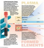
Plasma: The liquid matrix of blood, making up about 55% of its volume.
Formed Elements: Cells and cell fragments, including red blood cells (RBCs), white blood cells (WBCs), and platelets.

Functions of Blood
Transport: Carries oxygen, carbon dioxide, nutrients, hormones, and metabolic wastes.
Regulation: Maintains pH, ion composition, and body temperature.
Protection: Restricts fluid loss at injury sites, defends against toxins and pathogens.
Physical Characteristics of Blood
Temperature: 38ºC (100.4ºF)
Viscosity: High, due to formed elements and plasma proteins
pH: Slightly alkaline (7.35–7.45)
Volume: About 7% of body weight (e.g., 5.25 L in a 75-kg person)
Plasma
Composed of 92% water, 7% plasma proteins, and 1% other solutes (e.g., electrolytes, nutrients, gases).
Plasma proteins include albumins (osmotic pressure, transport), globulins (antibodies, transport), and fibrinogen (clotting).
Most plasma proteins are synthesized in the liver.
Formed Elements of Blood
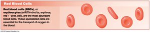
Red Blood Cells (Erythrocytes)
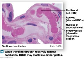
Red blood cells (RBCs) are the most abundant formed elements, responsible for oxygen transport. They are biconcave discs, which increases surface area for gas exchange and allows flexibility in capillaries.
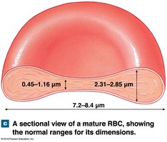
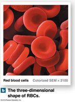
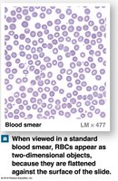
Structure: Biconcave, anucleate, lack mitochondria and ribosomes.
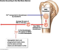
Lifespan: About 120 days.
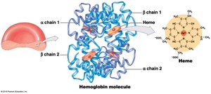
Hemoglobin: Each RBC contains about 280 million hemoglobin molecules, each capable of binding oxygen and carbon dioxide.
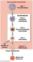
Red Blood Cell Production (Erythropoiesis)
Occurs in red bone marrow from hematopoietic stem cells (hemocytoblasts).
Stimulated by erythropoietin (EPO), mainly from the kidneys in response to hypoxia.
Stages: Myeloid stem cell → Proerythroblast → Erythroblast → Reticulocyte → Mature RBC.
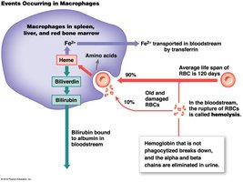
Hemoglobin Recycling
Old or damaged RBCs are phagocytized by macrophages in the spleen, liver, and bone marrow. Hemoglobin is broken down, and its components are recycled or excreted.
Iron: Recycled and transported by transferrin.
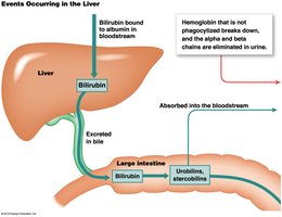
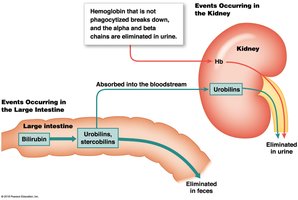
Heme: Converted to biliverdin, then bilirubin, which is excreted in bile.
Globin: Broken down into amino acids.
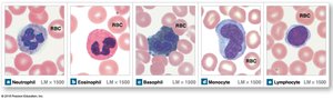
White Blood Cells (Leukocytes)
White blood cells are involved in the body's defense mechanisms. They are classified as granulocytes (neutrophils, eosinophils, basophils) and agranulocytes (monocytes, lymphocytes).
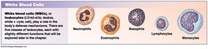
Neutrophils: Phagocytize bacteria, most abundant WBC.
Eosinophils: Attack parasites, involved in allergic reactions.
Basophils: Release histamine and heparin, involved in inflammation.
Monocytes: Become macrophages, phagocytize pathogens and debris.
Lymphocytes: Include T cells, B cells, and NK cells; involved in specific immunity.
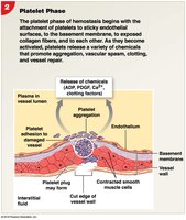
Platelets (Thrombocytes)
Platelets are small, membrane-bound cell fragments essential for blood clotting. They are produced in the bone marrow by megakaryocytes.
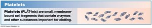
Circulate for 9–12 days, removed by the spleen.
Functions: Release clotting chemicals, form temporary plugs, reduce vessel break size.
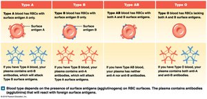
Blood Types and Transfusion Compatibility
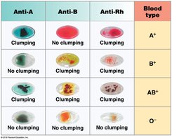
ABO and Rh Blood Groups
Blood type is determined by the presence or absence of specific surface antigens (agglutinogens) on RBCs: A, B, and Rh (D).
Type A: Surface antigen A, anti-B antibodies.
Type B: Surface antigen B, anti-A antibodies.
Type AB: Both antigens, no anti-A or anti-B antibodies.
Type O: No antigens, both anti-A and anti-B antibodies.
Rh+: Rh antigen present; Rh−: Rh antigen absent.
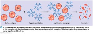
Transfusion Reactions and Compatibility Testing
If incompatible blood is transfused, antibodies in the recipient's plasma may cause agglutination and hemolysis of donor RBCs. Compatibility testing (cross-matching) is essential before transfusions.
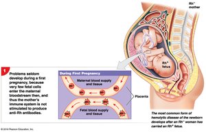
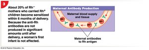
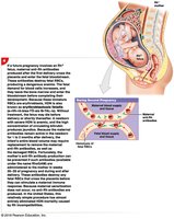
Hemolytic Disease of the Newborn (HDN)
HDN can occur when an Rh− mother carries an Rh+ fetus, leading to maternal antibody production against fetal RBCs in subsequent pregnancies.

Hemostasis and Blood Clotting
Phases of Hemostasis
Hemostasis is the process of stopping blood loss after injury and involves three phases:
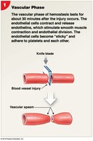
Vascular Phase: Vascular spasm constricts the vessel.
Platelet Phase: Platelets adhere to the injury site and form a plug.
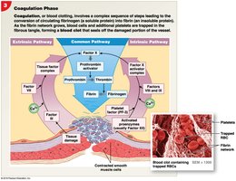
Coagulation Phase: Cascade of reactions leads to fibrin formation and blood clot.
Clot Retraction and Fibrinolysis
Clot Retraction: Platelets contract, pulling torn vessel edges together.
Fibrinolysis: Plasmin digests fibrin, dissolving the clot after repair.
Summary Table: Main Components of Blood
Component | Percentage | Main Functions |
|---|---|---|
Plasma | ~55% | Transport, osmotic balance, pH regulation |
Red Blood Cells | ~44% | Oxygen and carbon dioxide transport |
White Blood Cells | <1% | Immune defense |
Platelets | <1% | Clotting |
Additional info: This guide covers the essential concepts of blood structure, function, and clinical relevance for ANP college students, integrating textbook-level explanations and visual aids for comprehensive understanding.
