 Back
BackChapter 19: Blood – Structure, Function, and Clinical Significance
Study Guide - Smart Notes
 Tailored notes based on your materials, expanded with key definitions, examples, and context.
Blood and the Cardiovascular System
Overview of the Cardiovascular System
The cardiovascular system is essential for transporting substances throughout the body and maintaining homeostasis. It consists of three main components: blood, the heart, and blood vessels.
Blood: Specialized connective tissue containing cells suspended in a fluid matrix.
Heart: A muscular pump that circulates blood.
Blood Vessels: Conducting passageways that carry blood throughout the body.
Functions of Blood
Transport: Dissolved gases (O2, CO2), nutrients, hormones, and metabolic wastes.
Regulation: pH and ion composition of interstitial fluids.
Restriction: Limits fluid losses at injury sites.
Defense: Protects against toxins and pathogens.
Stabilization: Redistributes heat to maintain body temperature.
Characteristics of Blood
Temperature: 38°C (100.4°F)
Viscosity: 5 times thicker than water
pH: Slightly alkaline (7.35–7.45)
Volume: Approximately 7% of body weight in kilograms
Fractionation of Blood
Fractionation is the process of separating whole blood into plasma and formed elements.
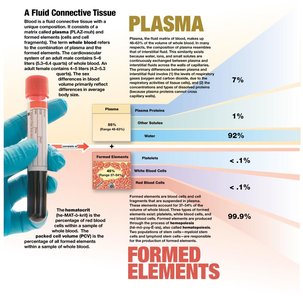
The Composition of Whole Blood
Plasma
Plasma is the fluid component of blood, making up 46–63% of blood volume. It consists primarily of water, plasma proteins, and other solutes.
Water: 92% of plasma
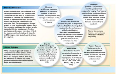
Plasma Proteins: Albumins, globulins, fibrinogen
Other Solutes: Organic nutrients, wastes, electrolytes
Plasma is similar to interstitial fluid but differs in protein and gas concentrations.
Formed Elements
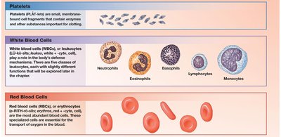
Formed elements are the cellular components of blood, including red blood cells, white blood cells, and platelets.
Red Blood Cells (RBCs): Erythrocytes, responsible for oxygen transport
White Blood Cells (WBCs): Leukocytes, involved in immune defense
Platelets: Cell fragments important for clotting
Red Blood Cells (RBCs)
Structure and Function
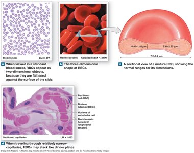
Red blood cells are highly specialized for oxygen transport. They are biconcave discs, which increases their surface-area-to-volume ratio and allows for efficient gas exchange.
Biconcave Shape: Facilitates absorption and release of oxygen
Rouleaux Formation: RBCs stack for smooth flow through capillaries
Anucleate: Lack nuclei, cannot divide or repair
No Mitochondria/Ribosomes: Rely on anaerobic metabolism
Hemoglobin
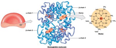
Hemoglobin is the protein responsible for oxygen and carbon dioxide transport in RBCs. It has a quaternary structure with four polypeptide chains, each containing a heme group.
Oxyhemoglobin (HbO2): Hemoglobin bound to oxygen
Deoxyhemoglobin: Hemoglobin not carrying oxygen
Carbaminohemoglobin: Hemoglobin bound to carbon dioxide
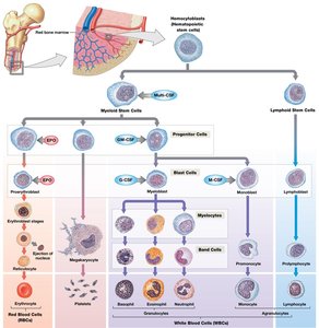
RBC Formation and Turnover
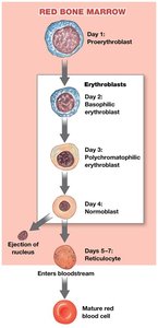
RBCs are produced through erythropoiesis in red bone marrow. They have a lifespan of about 120 days and are continuously replaced.
Hemocytoblasts: Stem cells that give rise to RBCs
Erythropoiesis: Process of RBC formation
Stages: Proerythroblast → Erythroblast → Normoblast → Reticulocyte → Mature RBC
Hemoglobin Recycling
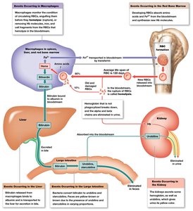
When RBCs are destroyed, their components are recycled. Iron is stored or transported, and heme is converted to biliverdin and then bilirubin, which is excreted in bile.
Hemolysis: RBC rupture
Macrophages: Phagocytize old RBCs
Bilirubin: Excreted by the liver; buildup causes jaundice
Iron: Transported by transferrin, stored as ferritin/hemosiderin
Blood Types and Cross-Reactions
Blood Type Determination
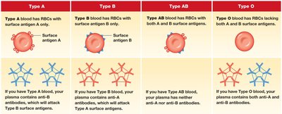
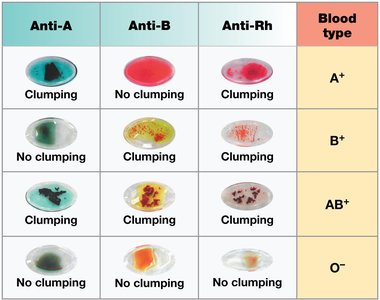
Blood type is determined by the presence or absence of surface antigens (agglutinogens) on RBCs. There are four main blood types: A, B, AB, and O.
Type A: Surface antigen A; anti-B antibodies
Type B: Surface antigen B; anti-A antibodies
Type AB: Both antigens; no anti-A or anti-B antibodies
Type O: No antigens; both anti-A and anti-B antibodies
Rh Blood Group
The Rh blood group is based on the presence or absence of the Rh (D) antigen. Rh+ individuals have the antigen, while Rh- individuals do not.
Rh+: Has Rh antigen; no anti-Rh antibodies
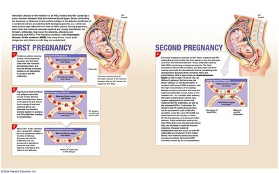
Rh-: Lacks Rh antigen; can develop anti-Rh antibodies if exposed
Hemolytic Disease of the Newborn: Occurs with Rh incompatibility between mother and fetus
Transfusion Reactions and Compatibility Testing
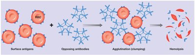
Transfusion reactions occur when incompatible blood types are mixed, leading to agglutination and hemolysis. Compatibility testing is essential before transfusions.
Cross-match Testing: Checks for reactions between donor and recipient
Blood Typing: Identifies A, B, and Rh antigens
Universal Donor: Type O-
White Blood Cells (WBCs)
Types and Functions
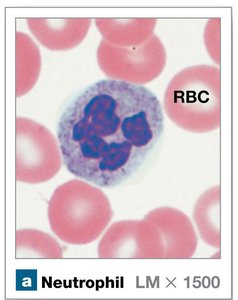
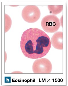
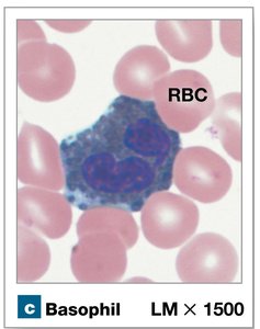
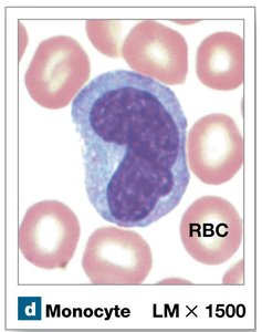
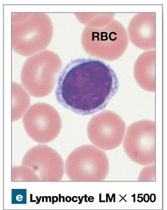
White blood cells, or leukocytes, are crucial for immune defense. They are classified as granular or agranular based on the presence of cytoplasmic granules.
Granular Leukocytes: Neutrophils, eosinophils, basophils
Agranular Leukocytes: Monocytes, lymphocytes
Classes of Lymphocytes
T Cells: Cell-mediated immunity
B Cells: Humoral immunity (antibody production)
Natural Killer (NK) Cells: Immune surveillance
WBC Production and Regulation
WBCs are produced from hemocytoblasts in bone marrow. Colony-stimulating factors (CSFs) regulate their production and differentiation.
Leukopoiesis: Formation of WBCs
Lymphocytopoiesis: Formation of lymphocytes
CSFs: Multi-CSF, GM-CSF, G-CSF, M-CSF
Platelets and Hemostasis
Platelets
Platelets are cell fragments essential for blood clotting. They are produced from megakaryocytes in bone marrow and circulate for 9–12 days.
Functions: Release clotting chemicals, patch vessel walls, reduce vessel break size
Thrombocytopoiesis: Platelet production
Regulation: Thrombopoietin, Interleukin-6, Multi-CSF
Hemostasis
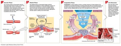
Hemostasis is the process of stopping bleeding, involving vascular, platelet, and coagulation phases.
Vascular Phase: Vascular spasm reduces blood flow
Platelet Phase: Platelet adhesion and aggregation form a plug
Coagulation Phase: Clotting factors activate pathways to form a blood clot
Clot Retraction: Stabilizes and repairs the vessel
Fibrinolysis: Gradual dissolution of the clot
Coagulation Pathways
Coagulation involves three pathways: extrinsic, intrinsic, and common. Each pathway activates clotting factors, leading to the conversion of fibrinogen to fibrin.
Extrinsic Pathway: Initiated by tissue factor from damaged cells
Intrinsic Pathway: Initiated by exposure to collagen and platelet factors
Common Pathway: Activation of Factor X, prothrombin to thrombin, fibrinogen to fibrin
Key Equation:
Regulation and Disorders
Anticoagulants: Inhibit clotting (e.g., antithrombin III, heparin, thrombomodulin)
Calcium and Vitamin K: Essential for clotting factor synthesis
Disorders: Thrombocytopenia, hemophilia, thrombophilia, deep vein thrombosis, pulmonary embolism
Summary Table: Blood Components
Component | Main Function | Key Features |
|---|---|---|
Plasma | Transport, regulation | 92% water, proteins, solutes |
RBCs | Oxygen transport | Biconcave, hemoglobin, anucleate |
WBCs | Immune defense | Granular/agranular, nucleated |
Platelets | Clotting | Cell fragments, short lifespan |
