 Back
BackChapter 20: The Lymphatic System and Immunity – Structured Study Notes
Study Guide - Smart Notes
 Tailored notes based on your materials, expanded with key definitions, examples, and context.
Structure and Function of the Lymphatic System
Introduction to the Immune and Lymphatic Systems
The immune and lymphatic systems work together to provide immunity, protecting the body from cellular injury and pathogens. The immune system consists of cells and proteins in blood and tissues, while the lymphatic system is a group of organs and tissues that also maintains fluid homeostasis.
Immune System: Includes leukocytes (white blood cells) and immune proteins in plasma.
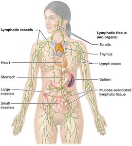
Lymphatic System: Composed of lymphatic vessels (blind-ended tubes) and lymphatic tissue/organs (tonsils, lymph nodes, spleen, thymus).
Functions of the Lymphatic System
The lymphatic system has three main functions:
Regulation of Interstitial Fluid Volume: Returns 2–4 liters of fluid lost from plasma daily to circulation, preventing drops in blood volume and pressure.
Absorption of Dietary Fats: Dietary fats enter lymphatic vessels (lacteals) in the small intestine and are delivered to the blood.
Immune Functions: Lymphoid organs filter pathogens from lymph and blood.
Lymphatic Vessels and Lymph Circulation
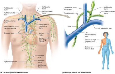
Lymphatic vessels collect lymph and merge to form lymph trunks, which drain specific body regions. The main trunks include lumbar, jugular, intestinal, bronchomediastinal, and subclavian trunks. The cisterna chyli is a large vessel that drains into the thoracic duct, which empties into the left internal jugular and subclavian veins. The right lymphatic duct drains the upper right side of the body.
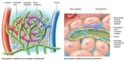
Lymphatic vessels are low-pressure circuits with valves to prevent backflow. Lymphatic capillaries are blind-ended, forming a one-way system. Their walls are leaky, allowing fluid and immune cells to enter.
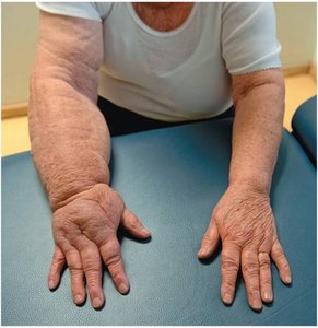
Lymphedema
Lymphedema is swelling caused by accumulation of excess interstitial fluid, often due to removal or blockage of lymphatic vessels. It prevents fluid return to the cardiovascular system, causing tissue enlargement.
Lymphoid Tissues and Organs
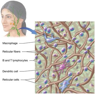
Reticular Tissue and Cells
Lymphatic tissue is a type of loose connective tissue called reticular tissue, containing specialized cells and reticular fibers that trap pathogens. Lymphoid organs house leukocytes, including macrophages, B and T lymphocytes, dendritic cells, and reticular cells.
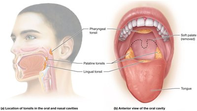
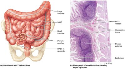
Mucosa-Associated Lymphatic Tissue (MALT)
MALT consists of clusters of lymphoid tissue protecting mucous membranes. Specialized MALT includes:
Tonsils: Pharyngeal, palatine, and lingual tonsils around oral and nasal cavities.
Peyer's Patches: Located in the ileum of the small intestine.
Appendix: Defends against bacteria in the large intestine.
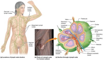
Lymph Nodes
Lymph nodes are bean-shaped clusters along lymphatic vessels, trapping pathogens in reticular "nets" and preventing their spread. They filter lymph and house immune cells.
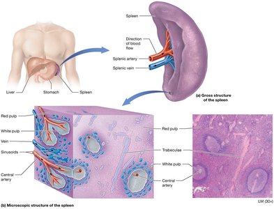
Spleen
The spleen is the largest lymphoid organ, filtering pathogens from blood and destroying old erythrocytes. It has two regions: red pulp (macrophages) and white pulp (leukocytes and dendritic cells).
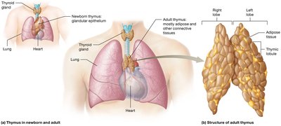
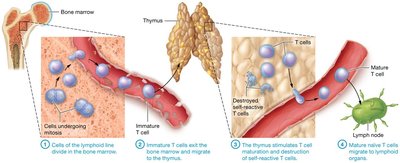
Thymus
The thymus generates functional T cells and is most active in children. It consists of lobules with cortex (dense T cells) and medulla (site of T cell destruction).
Overview of the Immune System
Lines of Defense
The immune system has three lines of defense:
First Line: Surface barriers (skin and mucous membranes).
Second Line: Innate immunity (cells and proteins).
Third Line: Adaptive immunity (cells and proteins).
Types of Immunity
Innate (Nonspecific) Immunity: Responds to all pathogens in the same way, using antimicrobial proteins and cells.
Adaptive (Specific) Immunity: Responds to unique antigens, involving cell-mediated (T cells) and antibody-mediated (B cells) immunity. Adaptive immunity has memory and is more efficient upon subsequent exposures.
Surface Barriers
Surface barriers block pathogen entry. Skin is resistant due to keratin and acidic sebum. Mucous membranes secrete mucus and acid to trap and destroy pathogens.
Cells and Proteins of Innate and Adaptive Immunity
Leukocytes: Agranulocytes (B and T lymphocytes, monocytes) and granulocytes (neutrophils, eosinophils, basophils).
Other Cells: NK cells, dendritic cells.
Proteins: Antibodies, complement system, cytokines.
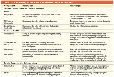
Innate Immunity: Internal Defenses
Cells of Innate Immunity
Phagocytes: Macrophages, neutrophils, eosinophils ingest pathogens.
Macrophages: First responders, kill pathogens, present antigens.
Neutrophils: Effective against bacteria, recruited to damaged tissues.
Dendritic Cells: Present antigens to T and B cells.
Eosinophils: Respond to parasitic pathogens.
NK Cells: Recognize and destroy cancerous and infected cells.
Basophils/Mast Cells: Mediate inflammation, involved in allergies.
Antimicrobial Proteins
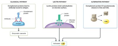
Complement System: 20+ plasma proteins activated by classical, lectin, or alternative pathways. Effects include cell lysis, enhanced inflammation, neutralization of viruses, opsonization, and clearance of immune complexes.
Cytokines
Tumor Necrosis Factor: Attracts and activates phagocytes.
Interferons: Inhibit viral replication.
Interleukins: Stimulate immune cell production and activation.
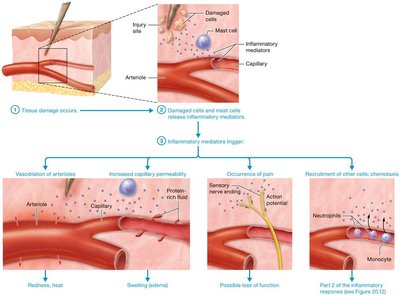
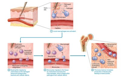
The Inflammatory Response
Inflammation occurs in response to cell damage. It involves release of mediators (histamine, cytokines), vasodilation, increased permeability, pain, and recruitment of leukocytes.
Anti-inflammatory Medications
NSAIDs: Inhibit cyclooxygenase, reducing prostaglandin production.
Corticosteroids: Inhibit formation of prostaglandins and leukotrienes.
Fever
Fever is an innate response to injury, triggered by pyrogens acting on the hypothalamus. It raises body temperature to enhance immune efficiency.
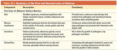
Summary Table: First and Second Lines of Defense
Component | Description | Function(s) |
|---|---|---|
Skin | Stratified squamous, keratinized epithelium | Physical barrier, resists trauma, secretes antimicrobial agents |
Mucous membranes | Epithelial membranes lining body passages | Trap pathogens, secrete mucus |
Phagocytes | Macrophages, neutrophils, eosinophils | Ingest pathogens and debris |
NK cells | Nonphagocytic lymphocytes | Destroy cancer and infected cells |
Complement | Plasma proteins | Cell lysis, inflammation, opsonization |
Cytokines | Signaling proteins | Regulate immune cell activity |
Fever | Elevated body temperature | Enhances phagocyte activity |
Adaptive Immunity: Cell-Mediated Immunity
T Cell Response
T Cells: Formed in bone marrow, mature in thymus. Clones respond to specific antigens.
Antigen: Substance recognized by B or T cells; immunogens generate immune response.
MHC Molecules: Class I (all nucleated cells, endogenous antigens), Class II (APCs, exogenous antigens).
T Cell Activation
Clonal Selection: Dendritic cells present antigens to T cells, activating specific clones.
Effector and Memory T Cells: Activated T cells proliferate and differentiate.
Effects of T Cells
Helper T Cells (TH): Secrete cytokines, activate macrophages, TC cells, and B cells.
Cytotoxic T Cells (TC): Kill infected, cancerous, or foreign cells by releasing perforin and enzymes.
Adaptive Immunity: Antibody-Mediated Immunity
B Cell Activation and Antibody Production
B Cells: Mature in bone marrow, recognize specific antigens.
Activation: B cell binds antigen, presents it to TH cell, differentiates into plasma and memory B cells.
Antibody Structure and Classes
Structure: Y-shaped molecule with two heavy and two light chains; constant and variable regions.
Classes: IgG, IgA, IgM, IgE, IgD (GAMED mnemonic).
Functions of Antibodies
Agglutination/Precipitation: Clumping of cells or molecules for easier phagocytosis.
Opsonization: Coating pathogens to enhance phagocytosis.
Neutralization: Binding toxins or viruses to prevent harm.
Complement Activation: IgM and IgG activate complement proteins.
Stimulation of Inflammation: IgE triggers release of mediators from mast cells and basophils.
Immunological Memory
Primary Response: Slow, initial exposure to antigen.
Secondary Response: Rapid, efficient response upon re-exposure, mainly IgG.
Vaccination: Induces memory cells for future protection.
Disorders of the Immune System
Hypersensitivity Disorders
Type I (Immediate): Allergies; IgE-mediated, rapid response, can cause anaphylactic shock.
Immunodeficiency Disorders
Primary: Genetic or developmental.
Secondary: Acquired (e.g., AIDS caused by HIV-1).
Autoimmune Disorders
Autoimmunity: Immune system attacks self antigens, leading to diseases like multiple sclerosis and type 1 diabetes.
Additional info: These notes expand on brief points with academic context, definitions, and examples for clarity and completeness.
