 Back
BackChapter 22: The Lymphatic System and Immunity – Structured Study Notes
Study Guide - Smart Notes
 Tailored notes based on your materials, expanded with key definitions, examples, and context.
The Lymphatic System and Immunity
Introduction to the Lymphatic and Immune Systems
The lymphatic system (also called the lymphoid system) consists of cells, tissues, and organs responsible for defending the body from environmental hazards and internal threats. The immune system includes all body cells and tissues involved in immunity, spanning the lymphatic, integumentary, skeletal, cardiovascular, respiratory, and digestive systems.
Immunity: The ability to resist infection and disease.
Pathogens: Disease-causing organisms such as viruses, bacteria, fungi, and parasites.

Components of the Lymphatic System
Main Components
The lymphatic system is composed of several key elements that work together to maintain immune function and fluid balance.
Lymph: Fluid similar to plasma but lacking plasma proteins.
Lymphatic vessels: Carry lymph from peripheral tissues to veins.
Lymphoid cells: Includes lymphocytes, phagocytes, and other immune cells.
Lymphoid tissues: Connective tissues dominated by lymphocytes.
Lymphoid organs: Sites where lymphocytes form, mature, or become activated.
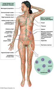
Primary and Secondary Lymphoid Tissues
Lymphoid tissues are classified based on their function in lymphocyte development and activation.
Primary lymphoid tissues and organs: Sites where lymphocytes are formed and mature (red bone marrow and thymus).
Secondary lymphoid tissues and organs: Sites where lymphocytes are activated (tonsils, MALT, lymph nodes, spleen).
Functions of the Lymphatic System
Production, maintenance, and distribution of lymphocytes and other lymphoid cells.
Return of excess tissue fluid to the bloodstream to maintain normal blood volume.
Regulation of interstitial fluid composition by transporting hormones, nutrients, and wastes.
Lymphatic Vessels and Meningeal Lymphatics
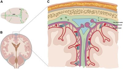
Lymphatic vessels (lymphatics) carry lymph from peripheral tissues to the venous system. Meningeal lymphatics are a network of vessels in the dura mater that drain cerebrospinal fluid (CSF) to cervical lymph nodes, remove wastes, and transport immune cells.
Lymphatic Capillaries and Lymph Flow
Lymphatic Capillaries
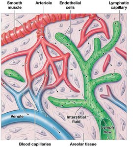
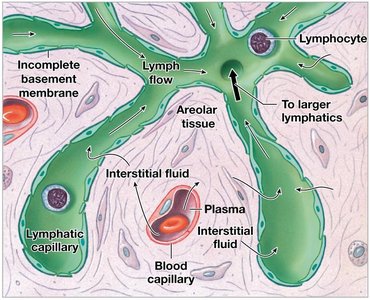
Lymphatic capillaries are the smallest lymphatic vessels, present in almost every tissue. Specialized capillaries called lacteals transport lipids absorbed by the digestive tract.
Closed at one end and begin in tissues.
Larger luminal diameters and thinner walls than blood capillaries.
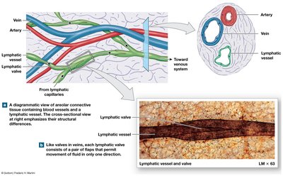
Endothelial cells overlap, forming one-way valves that allow entry of fluids, solutes, viruses, and bacteria, but prevent their return.
Lymphatic Vessels and Lymphedema
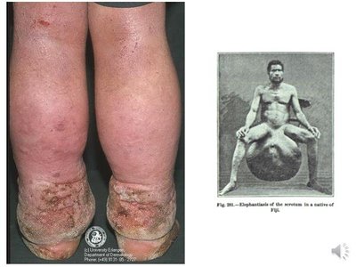
Lymph flows from capillaries to larger vessels, which have three layers similar to veins and valves to prevent backflow. Lymphedema is caused by obstruction of lymphatic vessels, leading to severe swelling and impaired immune function.
Major Lymph-Collecting Vessels
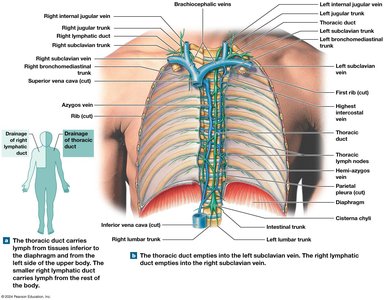
Superficial and deep lymphatics converge to form large lymphatic trunks, which empty into two major collecting vessels:
Thoracic duct: Collects lymph from all regions inferior to the diaphragm and the left side superior to the diaphragm; empties into the left subclavian vein.
Right lymphatic duct: Collects lymph from the right side superior to the diaphragm; empties into the right subclavian vein.
Lymphoid Cells, Tissues, and Organs
Lymphoid Cells
Lymphoid cells include immune system cells and support cells in lymphoid tissues.
Phagocytic cells: Macrophages, microphages, and other phagocytes.
Lymphocytes: T cells (thymus-dependent), B cells (bone marrow-derived), and NK (natural killer) cells.
Lymphoid Tissues and Nodules
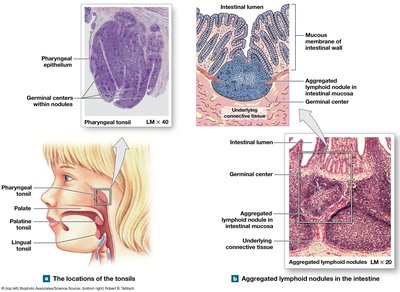
Lymphoid nodules are areolar tissue densely packed with lymphocytes. Tonsils are large lymphoid nodules in the pharynx. MALT (mucosa-associated lymphoid tissue) is found in the digestive, respiratory, urinary, and reproductive systems. Peyer's patches are clusters of lymphoid nodules deep to the intestinal epithelium.
Lymphoid Organs
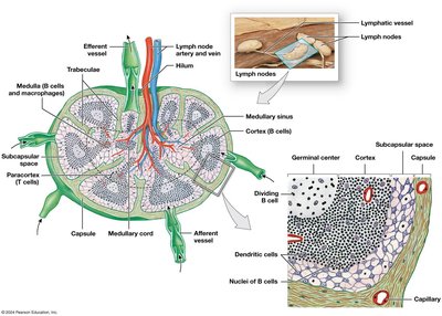
Lymphoid organs are separated from surrounding tissues by a connective tissue capsule. Lymph nodes are small organs found along lymphatic vessels, filtering lymph before it returns to the venous circulation.
Structure: Capsule, trabeculae, hilum, afferent and efferent lymphatics.
Lymph flow: Afferent lymphatics → subcapsular space → cortex (B cells) → paracortex (T cells) → medulla (B cells, macrophages) → efferent lymphatics at hilum.
Lymph Node Functions and Disorders
Filters lymph, removes 99% of antigens.
Phagocytes engulf debris and pathogens.
Monitors peripheral infections and activates immune response.
Largest lymph nodes are in groin, axillae, and base of neck.
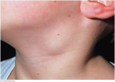
Lymphadenitis: Inflammation of lymph nodes.
Lymphadenopathy: Chronic or excessive enlargement of lymph nodes.
Thymus
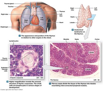
The thymus is a pink, grainy lymphoid organ located in the mediastinum above the heart. It atrophies after puberty and becomes inactive.
Divided into two lobes, each with cortex (densely packed lymphocytes) and medulla (epithelial reticular cells in Hassall's corpuscles).
Regulates T cell development and maturation.
Produces thymosin, a hormone promoting T cell maturation.
Spleen
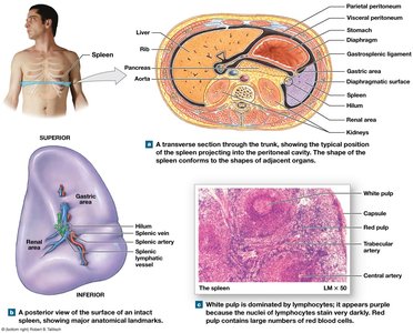
The spleen is a large lymphoid organ lateral to the stomach. It filters blood, removes abnormal blood cells, stores iron, and mounts immune responses to antigens in blood.
Red pulp: Contains many red blood cells.
White pulp: Resembles lymphoid nodules.
Rupture can cause internal bleeding; splenectomy increases risk of bacterial infections.
Innate and Adaptive Immunity
Types of Immunity
Immunity is the ability to resist and defend against infectious organisms and other damaging substances. The immune response is the body's reaction to infectious agents and abnormal substances.
Innate (nonspecific) immunity: Physical barriers and internal defenses present at birth; does not distinguish threats.
Adaptive (specific) immunity: Protects against specific antigens; depends on B and T lymphocytes; develops after exposure.
Lymphocyte Production and Distribution
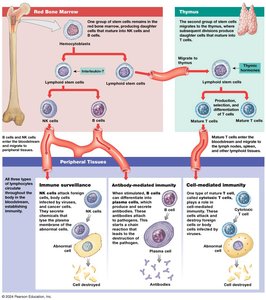
Lymphocytes (B cells, T cells, NK cells) are produced in the red bone marrow, thymus, and peripheral lymphoid tissues. Hemocytoblasts in bone marrow divide to produce lymphoid stem cells, which differentiate into all lymphocytes.
Innate Defenses
Physical Barriers
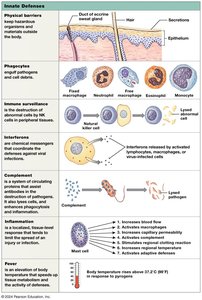
Physical barriers prevent pathogens from entering the body. These include skin, hair, mucous membranes, mucus, sweat, urine, and secretions containing enzymes, antibodies, or stomach acid.
Phagocytes
Phagocytes attack and engulf microorganisms and debris. Types include microphages (neutrophils, eosinophils) and macrophages (derived from monocytes).
Fixed macrophages: Reside in specific tissues (e.g., microglia in CNS, Kupffer cells in liver).
Free macrophages: Travel throughout the body (e.g., alveolar macrophages in lungs).
Phagocytosis begins with adhesion, followed by internalization and digestion.
Immune Surveillance
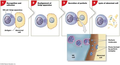
Natural killer (NK) cells look for abnormal cells in peripheral tissues, such as cancer cells or virus-infected cells. NK cell activation involves recognition, realignment of the Golgi apparatus, secretion of perforins, and lysis of the abnormal cell.
Interferons
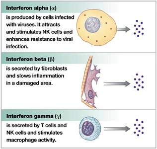
Interferons (IFNs) are small proteins released by activated lymphocytes, macrophages, and virus-infected tissue cells. They are cytokines that trigger production of antiviral proteins in healthy cells.
Complement System
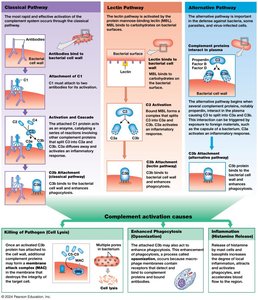
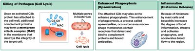
The complement system consists of more than 30 special proteins in plasma that assist antibodies in destroying pathogens. Activation occurs via classical, lectin, or alternative pathways, all leading to conversion of C3 to C3a and C3b.
Inflammation and Fever
Inflammation is a localized tissue response to injury, characterized by redness, swelling, heat, and pain. It repairs injury, slows pathogen spread, and mobilizes defenses. Fever is an elevated body temperature induced by pyrogens, increasing metabolic rate and inhibiting some pathogens.
Adaptive Defenses
T Cells and Immunity
Adaptive defenses result from coordinated activities of T cells and B cells. T cells include cytotoxic, helper, regulatory, memory, inflammatory, and suppressor-inducer types.
Cell-mediated immunity: Mediated by cytotoxic T cells; defends against abnormal cells and pathogens inside cells.
Antibody-mediated immunity: Mediated by B cells; defends against antigens and pathogens in body fluids.
Antigen Presentation and Recognition
T cells recognize antigens presented by antigen-presenting cells (APCs) via MHC proteins. Class I MHC proteins are found on all nucleated cells; Class II MHC proteins are on lymphocytes and APCs. CD markers (CD3, CD4, CD8) facilitate antigen recognition.
B Cells and Immunity
B Cell Sensitization and Activation
B cells have specific surface antibodies (B cell receptors). When an antigen binds, it is taken in, processed, and presented on a class II MHC protein. Helper T cells secrete cytokines to promote B cell activation, leading to plasma cell formation and antibody secretion. Memory B cells remain in reserve.
Antibody Structure and Function
Antibodies are soluble proteins composed of two pairs of polypeptide chains (heavy and light), each with constant and variable segments. The variable segments form antigen-binding sites.
Classes of Antibodies (Immunoglobulins)
IgG: Most abundant; crosses placenta; provides passive immunity to fetus.
IgE: Attaches to basophils and mast cells; important in allergic response.
IgD: Found on B cells; involved in sensitization.
IgM: First antibody secreted; responsible for agglutination of incompatible blood types.
IgA: Found in glandular secretions; attacks pathogens before they access internal tissues.
Actions of Antibodies
Neutralization
Precipitation and agglutination
Activation of complement system
Attraction of phagocytes
Opsonization
Stimulation of inflammation
Prevention of bacterial and viral adhesion
Immune Responses to Antigen Exposure
Primary response: Slow development; IgM produced first, then IgG.
Secondary response: Rapid and extensive; memory cells activate quickly; IgG rises rapidly.
Immune Responses to Infection
Bacterial Infection
Neutrophils and NK cells migrate to the area, cytokines draw more phagocytes, cytotoxic T cells destroy bacteria, B cells produce antibodies, and antibodies target bacteria for destruction.
Viral Infection
Infected cells release interferons, present viral proteins to activate NK and cytotoxic T cells, APCs activate helper T cells, B cells produce antibodies to target the virus.
Immune Disorders and Effects of Aging
Stress and Immune Response
Chronic stress increases glucocorticoid secretion, which depresses inflammation, reduces phagocyte activity, and inhibits interleukin secretion, lowering resistance to disease.
Immune Disorders
Hypersensitivities (allergies): Excessive immune responses to antigens; can cause anaphylaxis.
Autoimmune disorders: Immune system attacks self-antigens (e.g., thyroiditis, rheumatoid arthritis, type 1 diabetes, multiple sclerosis).
Immunodeficiency diseases: Result from developmental problems, viral infections, or immunosuppressive treatments (e.g., SCID, HIV/AIDS).
Effects of Aging on Immune Response
Reduced thymic hormone production.
T cells less responsive to antigens.
Fewer helper T cells reduce B cell responsiveness.
Decline in immune surveillance against tumor cells.
Integration of the Lymphatic System
The lymphatic system integrates with other body systems to maintain homeostasis and defend against pathogens.
Table: Cells that Participate in Tissue Defenses
Cell | Functions |
|---|---|
Neutrophils | Phagocytosis; stimulation of inflammation |
Eosinophils | Phagocytosis of antigen–antibody complexes; suppression of inflammation; participation in allergic response |
Mast cells and basophils | Stimulation and coordination of inflammation by release of histamine, heparin, leukotrienes, prostaglandins |
Macrophages (free and fixed, stellate, microglia) | Phagocytosis; antigen processing; antigen presentation with class II MHC proteins; secretion of cytokines |
Dendritic cells | Pinocytosis; antigen processing; antigen presentation bound to class II MHC proteins |
NK cells | Destruction of plasma membranes containing abnormal antigens |
Cytotoxic T cells (CD8 marker) | Lysis of plasma membranes containing antigens bound to class I MHC proteins; secretion of perforins, defensins, lymphotoxins, and other cytokines |
Key Equations and Concepts
Antibody Titer: Level of antibodies in plasma.
Complement Activation:
Fever Threshold:
