Back
BackChapter 22: The Respiratory System – Structure and Function
Study Guide - Smart Notes
 Tailored notes based on your materials, expanded with key definitions, examples, and context.
Respiratory System Overview
Introduction to Respiration
The respiratory system is responsible for the exchange of gases between the atmosphere and the body’s cells. This process supports cellular metabolism and ATP production, which are essential for life.
Ventilation: Movement of air into and out of the lungs.
Gas Exchange: Oxygen (O2) moves from air to blood to tissues; carbon dioxide (CO2) moves from tissues to blood to lungs for exhalation.
Cellular Respiration: Cells use O2 to produce ATP, releasing CO2 as a waste product.
Organs of the Respiratory System
Major Structures
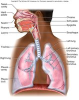
The respiratory system consists of the nose, pharynx, larynx, trachea, bronchi, and lungs. It is divided into upper and lower respiratory tracts. The diaphragm forms the floor of the thoracic cavity and is essential for breathing.
Upper Respiratory Tract
Nose: Structure and Function
The nose serves as the primary entryway for air. It moistens, warms, filters, and cleans inhaled air, detects odors, and acts as a resonating chamber for the voice.
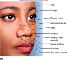
Anterior nares (nostrils): External openings for air entry.
Nasal septum: Divides the nasal cavity into right and left chambers.
Bridge and Apex: Structural features of the external nose.
Anatomy of the Nasal Region
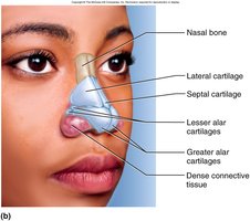
The nose is supported by both bone and cartilage. The superior half is formed by the nasal bones and maxillae, while the inferior half is shaped by various cartilages and dense connective tissue.
Nasal Cavity
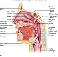
The nasal cavity extends from the nostrils to the posterior nares. It contains the vestibule (lined with stratified squamous epithelium and guard hairs), and the nasal conchae (superior, middle, inferior) which increase surface area and turbulence for air filtration. The nasal septum divides the cavity into right and left nasal fossae.
Nasal Cavity Mucosa
The nasal cavity is lined by two types of mucosa:
Olfactory mucosa: Located in the roof of the nasal fossa; detects odors.
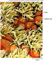
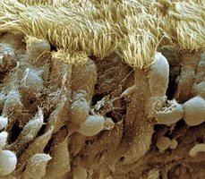
Respiratory mucosa: Ciliated pseudostratified epithelium that lines the rest of the nasal cavity; contains goblet cells that secrete mucus to trap particles, and lysozyme to destroy bacteria.
Cilia and Erectile Tissue in the Nasal Cavity
Cilia in the respiratory epithelium sweep mucus and trapped debris toward the pharynx to be swallowed. The erectile tissue of the inferior concha contains a venous plexus that periodically engorges with blood, shifting airflow and preventing drying of the mucosa. Trauma to this area can cause spontaneous epistaxis (nosebleed).
Pharynx
Regions of the Pharynx
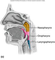
The pharynx is a muscular tube that serves as a passageway for air, food, and drink. It is divided into three regions:
Nasopharynx: Receives air from the nasal cavity; contains the pharyngeal tonsil and auditory tubes.
Oropharynx: Passage for air, food, and drink; contains palatine and lingual tonsils.
Laryngopharynx: Located at the level of the hyoid bone; passage for air, food, and drink.
Larynx (Voice Box)
Structure and Function
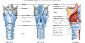
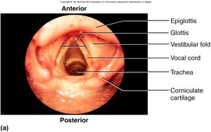
The larynx is a 4 cm cartilaginous chamber that keeps food and drink out of the airway and is essential for sound production. The epiglottis guards the glottis and directs food to the esophagus. The glottis consists of the vocal cords and the opening to the trachea. The larynx contains nine cartilages and muscular walls for speech control.
Infant larynx: Positioned higher in the throat, allowing simultaneous breathing and swallowing; descends by age 2.
Action of Vocal Cords
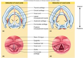
The vocal cords can be adducted (tightened for high-pitched sounds) or abducted (loosened for low-pitched sounds). Sound is produced by vibration of the cords, but speech requires additional modification by the pharynx, oral cavity, tongue, and lips.
Lower Respiratory Tract
Trachea (Windpipe)
The trachea is a rigid tube about 4.5 inches long and 2.5 cm in diameter, located anterior to the esophagus. It is supported by 16–20 C-shaped cartilaginous rings and lined with ciliated pseudostratified epithelium, which functions as a mucociliary escalator to move mucus and debris upward toward the pharynx.
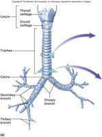
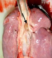
Tracheal Structure
The C-shaped cartilage rings keep the trachea open, while the trachealis muscle allows for diameter adjustment during coughing or swallowing.
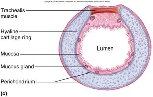
Tracheotomy and Tracheostomy
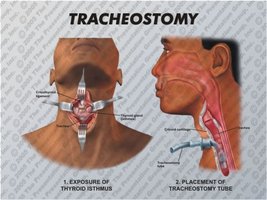
A tracheotomy is a surgical procedure to create an opening (tracheostomy) in the trachea to bypass upper airway obstructions. Air entering through a tracheostomy must be filtered and humidified.
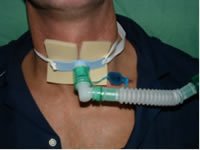
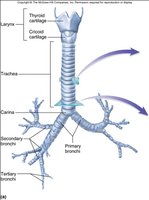
Carina and Bronchi
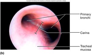
The carina is a median ridge at the point where the trachea divides into the right and left primary bronchi. This area is highly sensitive and triggers coughing if stimulated.
Lungs
Surface Anatomy
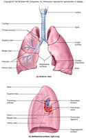
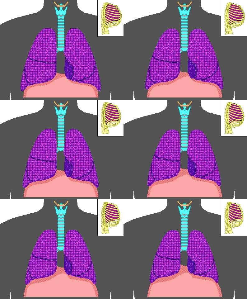
The lungs are paired organs with distinct lobes and surfaces. The right lung is shorter (due to the liver) and has three lobes, while the left lung is narrower (due to the heart) and has two lobes. The hilum is the entry/exit point for bronchi, blood vessels, and nerves.
Bronchial Tree
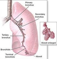
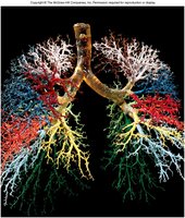
The bronchial tree consists of branching airways:
Primary bronchi: Right is wider and more vertical (higher risk of aspiration).
Secondary bronchi: Two in the left lung, three in the right lung.
Tertiary bronchi: Ten in the right lung, eight in the left lung; each supplies a bronchopulmonary segment.
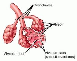
Bronchioles: Smaller branches leading to terminal bronchioles and alveolar sacs (sites of gas exchange).
Alveoli
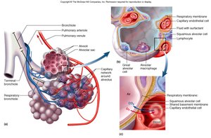
Alveoli are tiny air sacs where gas exchange occurs. Each alveolus is lined by:
Type I (Squamous) alveolar cells: Thin cells for gas exchange (95% of surface area).
Type II (Great) alveolar cells: Cuboidal cells that secrete surfactant to lower surface tension and prevent alveolar collapse.
Alveolar macrophages (dust cells): Immune cells that remove debris and pathogens.
Alveolar Blood Supply
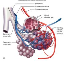
Alveoli are surrounded by a dense capillary network, facilitating efficient gas exchange between air and blood.
Pleurae and Pleural Fluid
Structure and Function
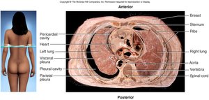
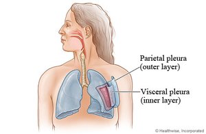
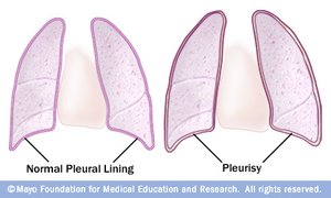
The lungs are enclosed by two layers of pleura:
Visceral pleura: Covers the lungs.
Parietal pleura: Lines the rib cage.
Pleural cavity: Space between pleurae, filled with lubricating fluid.
Functions include reducing friction, creating a pressure gradient for lung inflation, and compartmentalizing the thoracic cavity to prevent infection spread.
Pulmonary Ventilation (Breathing)
Mechanics of Breathing
Pulmonary ventilation consists of inspiration (inhalation) and expiration (exhalation). Quiet respiration occurs at rest, while forced respiration occurs during exercise. Airflow depends on pressure differences between the lungs and the atmosphere.
Respiratory Muscles
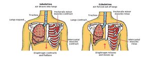
The diaphragm and external intercostal muscles expand the thoracic cavity during inspiration, decreasing pressure and drawing air in. Expiration is usually passive (elastic recoil), but can be forced by abdominal and internal intercostal muscles.
Neural Control of Breathing
Central Regulation
Breathing is controlled by repetitive neural stimuli from the brain, specifically the medulla oblongata and pons, which regulate unconscious breathing patterns.