 Back
BackChapter 22: The Respiratory System – Structure and Function
Study Guide - Smart Notes
 Tailored notes based on your materials, expanded with key definitions, examples, and context.
Overview of the Respiratory System
Major Functions and Processes
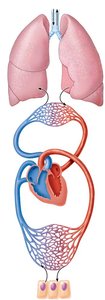
The respiratory system is essential for gas exchange, supplying oxygen to tissues and removing carbon dioxide. It works closely with the cardiovascular system to maintain homeostasis. The four basic processes of respiration are:
Pulmonary ventilation (breathing): Movement of air into and out of the lungs.
Pulmonary gas exchange: Exchange of gases between alveoli and blood.
Gas transport: Movement of gases in the blood between lungs and tissues.
Tissue gas exchange: Exchange of gases between blood and body tissues.
Additional functions include regulation of blood pH, voice production, protection against pathogens, and olfaction (smell).
Functional Anatomy of the Respiratory System
Regions of the Respiratory System
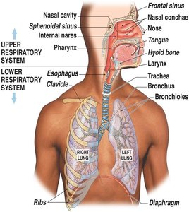
The respiratory system is divided into two main regions:
Upper respiratory tract: Nose, nasal cavity, paranasal sinuses, pharynx
Lower respiratory tract: Larynx, trachea, bronchi, bronchioles, alveoli
Upper Respiratory System
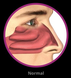
Nose and Nasal Cavity
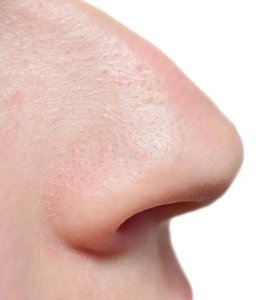
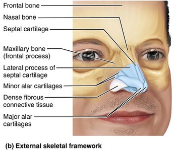
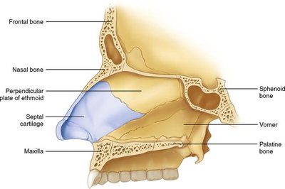
The nose is the only external part of the respiratory system. Its framework is formed by the frontal bone, paired nasal bones, maxilla, and hyaline cartilage. The nostrils (nares) allow air entry.
Functions: Provides airway, moistens/warms air, filters/cleans air, resonating chamber for speech, houses olfactory receptors.
Nasal Cavity Structure
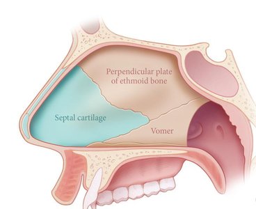
The nasal cavity extends from the external nares to the internal nares (choanae), divided by the nasal septum (ethmoid bone, vomer, and cartilage). The roof is formed by the ethmoid and sphenoid bones; the floor by the palatine bone and maxilla.
Mucosa of the Nasal Cavity
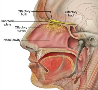
Olfactory mucosa: Contains olfactory receptors for smell.
Respiratory mucosa: Pseudostratified ciliated columnar epithelium with goblet cells; warms, humidifies, and traps debris/pathogens.
Nasal Conchae and Meatuses
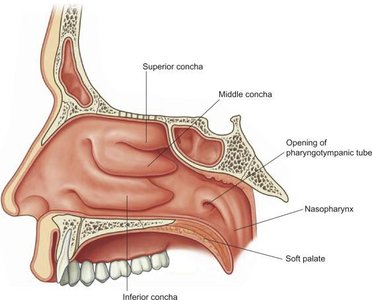
The lateral walls of the nasal cavity have three scroll-like projections (superior, middle, inferior conchae) with meatuses in between. These structures increase surface area, warm/humidify air, and create turbulence to trap debris.
Paranasal Sinuses
The paranasal sinuses are mucus-lined cavities in the frontal, sphenoid, ethmoid, and maxillary bones. They lighten the skull and help warm/moisten air.
Homeostatic Imbalances
Rhinitis: Inflammation of the nasal mucosa (caused by viruses, bacteria, or allergies).
Sinusitis: Inflammation of the paranasal sinuses, often following rhinitis; can cause sinus headaches.
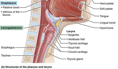
Pharynx
The pharynx is a funnel-shaped passageway connecting the nasal cavity and mouth to the larynx and esophagus. It consists of three regions:
Nasopharynx: Posterior to nasal cavity; passageway for air only; contains pharyngeal tonsil.
Oropharynx: Posterior to oral cavity; passageway for air and food; contains palatine and lingual tonsils.
Laryngopharynx: Posterior to larynx; passageway for air and food.
Lower Respiratory System
Conducting vs. Respiratory Zones
Conducting zone: Conduits for air (nose to terminal bronchioles); warm, humidify, and filter air.
Respiratory zone: Site of gas exchange (respiratory bronchioles, alveolar ducts, alveoli).
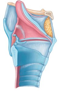
Larynx (Voice Box)
The larynx provides an open airway, routes air and food, and produces voice. It is composed of nine cartilages (mainly hyaline), including the thyroid cartilage (Adam's apple), cricoid cartilage, and epiglottis (elastic cartilage).
Epiglottis: Covers the laryngeal inlet during swallowing to prevent food entry into the trachea.
Vocal folds (true vocal cords): Vibrate to produce sound; flanked by vestibular folds (false vocal cords).
Trachea (Windpipe)
The trachea descends from the larynx and splits into two main bronchi. Its wall consists of mucosa (ciliated pseudostratified epithelium), submucosa (connective tissue with glands), and adventitia (outer connective tissue). The wall is reinforced by C-shaped hyaline cartilage rings, allowing esophageal expansion.
Bronchi and Bronchial Tree
The bronchial tree branches from the main bronchi into secondary (lobar) bronchi, tertiary (segmental) bronchi, and smaller bronchioles, ending with terminal bronchioles (end of conducting zone).
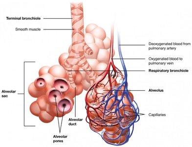
Respiratory Zone Structures
Terminal bronchioles lead to respiratory bronchioles, which lead to alveolar ducts and alveolar sacs (clusters of alveoli). The alveoli are the main site of pulmonary gas exchange.
Type I alveolar cells: Simple squamous epithelial cells for gas exchange.
Type II alveolar cells: Secrete surfactant to reduce surface tension and prevent alveolar collapse.
Alveolar macrophages: Remove debris and pathogens.
The respiratory membrane consists of the alveolar epithelium and capillary endothelium, across which gases diffuse.
Practice Questions
List the four types of respiration in order: Pulmonary ventilation, pulmonary gas exchange, gas transport, tissue gas exchange.
Which bones make up the external nose? (Maxilla, nasal bones, frontal bone; NOT mandible)
Which is NOT a function of the respiratory system? (Gustation)
Which structure seals the larynx during swallowing? (Epiglottis)
Which laryngeal cartilage forms the Adam’s apple? (Thyroid cartilage)
Where can gas exchange occur? (Respiratory bronchioles, not trachea or terminal bronchioles)
Alveoli are composed of what type of epithelium? (Simple squamous epithelium)
