 Back
BackChapter 22: The Respiratory System – Structure and Function
Study Guide - Smart Notes
 Tailored notes based on your materials, expanded with key definitions, examples, and context.
The Respiratory System: Overview and Functions
Introduction
The respiratory system is essential for gas exchange, speech, olfaction, and maintaining homeostasis. It consists of organs and structures that facilitate the movement of air and the exchange of oxygen (O2) and carbon dioxide (CO2) between the atmosphere and the bloodstream.
Gas Exchange: O2 is absorbed and CO2 is expelled between blood and air.
Speech and Vocalization: Air movement enables sound production.
Olfaction: The sense of smell is mediated by olfactory receptors in the nasal cavity.
pH Regulation: Removal of CO2 helps control blood pH.
Blood Pressure Regulation: The lungs produce angiotensin II, a vasoconstrictor.
Assists Circulation: Breathing aids venous and lymphatic return to the heart.
Anatomy of the Respiratory System
Major Structures
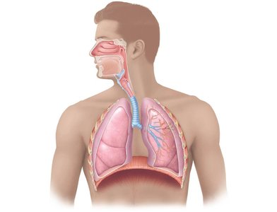
The respiratory system is divided into the upper and lower tracts, each with specialized structures for air conduction and gas exchange.
Upper Respiratory Tract: Includes the nose, nasal cavity, paranasal sinuses, and pharynx.
Lower Respiratory Tract: Includes the larynx, trachea, bronchi, bronchioles, and lungs.
The Nose and Nasal Cavity
The nose provides an airway, moistens and warms air, filters particles, resonates sound, and houses olfactory receptors. The nasal cavity contains conchae (turbinate bones) that increase surface area and enhance air conditioning.
Olfactory Mucosa: Contains smell receptors.
Respiratory Mucosa: Filters, heats, and moistens air.
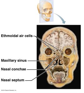
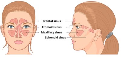
Paranasal Sinuses
Paranasal sinuses are air-filled spaces in the skull that open into the nasal cavity. They are lined by respiratory mucosa and help lighten the skull, warm and moisten air, and enhance voice resonance.
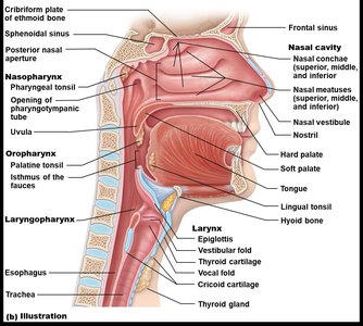
The Pharynx
The pharynx is a muscular tube that serves as a passageway for air and food. It is divided into three regions:
Nasopharynx: Lined with pseudostratified ciliated columnar epithelium; contains the pharyngeal tonsil and pharyngotympanic tube.
Oropharynx: Lined with nonkeratinized stratified squamous epithelium; contains palatine and lingual tonsils.
Laryngopharynx: Also lined with nonkeratinized stratified squamous epithelium; continuous with the esophagus and larynx.

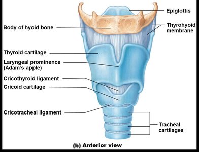
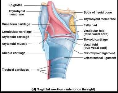
The Larynx
The larynx connects the pharynx to the trachea and is responsible for voice production, maintaining an open airway, and routing air and food using the epiglottis.
Cartilages: Includes thyroid, cricoid, arytenoid, and epiglottic cartilages.
Vocal Folds: True vocal cords produce sound; vestibular folds (false vocal cords) do not.
Lower Respiratory Tract
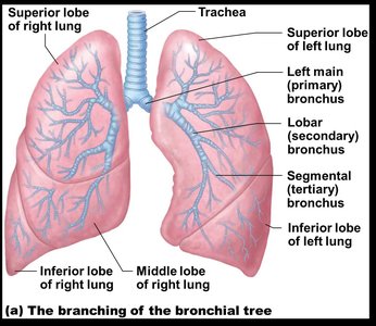
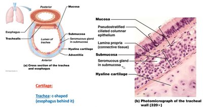
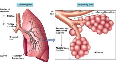
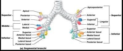
Trachea and Bronchial Tree
The trachea is a flexible tube supported by C-shaped hyaline cartilage rings. It divides into the right and left main (primary) bronchi, which further branch into secondary (lobar) and tertiary (segmental) bronchi, and then into bronchioles and terminal bronchioles.
Trachea: Lined with pseudostratified ciliated columnar epithelium; cartilage prevents collapse.
Bronchi: Right main bronchus is wider and more vertical than the left.
Bronchioles: Lack cartilage; smooth muscle controls airflow.
Conducting Zone vs. Respiratory Zone
The conducting zone includes all respiratory passages that carry air to the sites of gas exchange but do not participate in gas exchange themselves. The respiratory zone is where gas exchange occurs, beginning with respiratory bronchioles and including alveolar ducts and sacs.
Conducting Zone: Nasal cavity to terminal bronchioles; "anatomical dead space".
Respiratory Zone: Respiratory bronchioles, alveolar ducts, and alveoli.
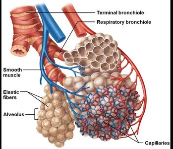
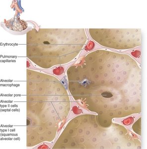
Alveoli and Gas Exchange
Structure of Alveoli
Alveoli are tiny air sacs where gas exchange occurs. They are surrounded by capillaries and have thin walls to facilitate diffusion.
Type I Alveolar Cells: Simple squamous epithelial cells forming the respiratory membrane.
Type II Alveolar Cells: Cuboidal cells that secrete surfactant to reduce surface tension.
Alveolar Macrophages: Remove debris and pathogens.
Alveolar Pores: Equalize air pressure and provide alternate air routes.
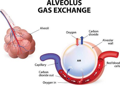
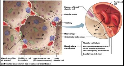
Gas Exchange Mechanism
Gas exchange occurs by diffusion across the respiratory membrane. Oxygen moves from alveoli into blood, while carbon dioxide moves from blood into alveoli to be exhaled.
Respiratory Membrane: Consists of alveolar epithelium, fused basement membrane, and capillary endothelium.
Surfactant: Reduces surface tension, preventing alveolar collapse.
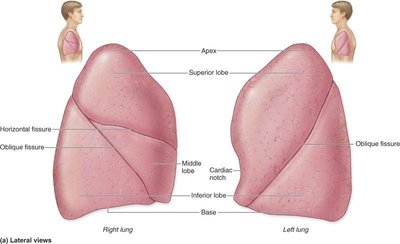
Lungs and Pleurae
Lung Anatomy
The lungs are divided into lobes and segments, each supplied by its own bronchus and blood vessels. The right lung has three lobes; the left lung has two lobes and a cardiac notch.
Bronchopulmonary Segments: Functionally independent units of the lung.
Pleurae: Double-layered serous membranes (parietal and visceral) that reduce friction and compartmentalize the lungs.
Respiratory Physiology and Terminology
Key Terms
Respiration: Overall process of gas exchange in the body.
External Respiration: Gas exchange between air in the lungs and blood.
Internal Respiration: Gas exchange between blood and tissues.
Pulmonary Ventilation: Movement of air into (inspiration) and out of (expiration) the lungs.
Cellular Respiration: Metabolic processes (glycolysis, Krebs cycle, oxidative phosphorylation) that produce ATP.
CO2 Transport and pH Regulation
CO2 is transported in the blood as dissolved gas, carbaminohemoglobin, and bicarbonate ion. The following equation summarizes the reversible reaction catalyzed by carbonic anhydrase:
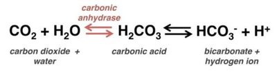
This reaction is crucial for maintaining acid-base balance in the blood.
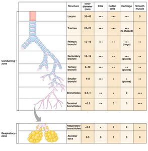
Summary Table: Conducting vs. Respiratory Zones
Structure | Inner Diameter (mm) | Cilia | Goblet Cells | Cartilage | Smooth Muscle |
|---|---|---|---|---|---|
Larynx | 35–45 | +++ | +++ | +++ (plates) | + |
Trachea | 20–25 | +++ | +++ | +++ (C-shaped) | + |
Primary Bronchi | 12–16 | +++ | +++ | +++ (rings) | + |
Secondary Bronchi | 8–10 | +++ | +++ | ++ (plates) | ++ |
Tertiary Bronchi | 1–8 | +++ | +++ | + (plates) | +++ |
Smaller Bronchi | 1–8 | ++ | ++ | + (plates) | +++ |
Terminal Bronchioles | <0.5 | + | 0 | 0 | +++ |
Respiratory Bronchioles | <0.5 | 0 | 0 | 0 | ++ |
Alveolar Sacs | 0 | 0 | 0 | 0 | 0 |
Additional info: The number of plus signs (+) indicates the relative abundance or presence of each feature.
Key Concepts and Clinical Relevance
Obstruction or damage to any part of the respiratory tract can impair gas exchange and lead to respiratory distress.
Surfactant deficiency (e.g., in premature infants) can cause alveolar collapse (atelectasis).
Segmental anatomy of the lungs is important for surgical resection and disease localization.
