 Back
BackChapter 22: The Respiratory System – Structure, Histology, and Function
Study Guide - Smart Notes
 Tailored notes based on your materials, expanded with key definitions, examples, and context.
Respiratory System Overview
Introduction
The respiratory system is responsible for the exchange of gases (oxygen and carbon dioxide) between the body and the environment. It consists of anatomical structures that conduct air into the lungs and facilitate gas exchange at the alveolar level.
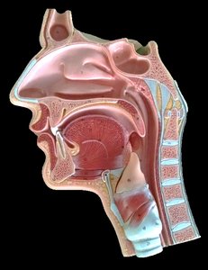
External Features of the Nose
Anatomical Landmarks
Alae: The lateral, flared portions of the nostrils.
Nares: The external openings of the nasal cavity (nostrils).
Philtrum: The vertical groove between the base of the nose and the upper lip.
Respiratory Spaces & Borders
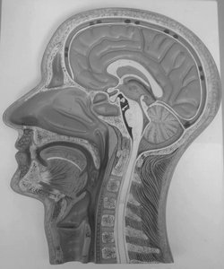
Nasal Cavity and Pharynx
Nasal Vestibule: The anterior part of the nasal cavity, just inside the nostrils.
Hard Palate: The bony anterior portion of the roof of the mouth, separating the nasal and oral cavities.
Soft Palate: The muscular posterior portion of the roof of the mouth.
Pharynx: A muscular tube that serves as a passageway for both air and food; divided into nasopharynx, oropharynx, and laryngopharynx.
Uvula: A small projection from the posterior edge of the soft palate.
Nasal Cavity – Conchae & Meatuses
Structure and Function
Conchae: Three bony projections (superior, middle, inferior) on the lateral walls of the nasal cavity that increase surface area and help warm, moisten, and filter air.
Meatuses: Passages beneath each concha that direct airflow and drain sinuses.
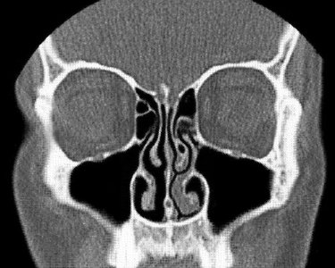
Paranasal Sinuses
Sinus Anatomy
Frontal Sinus: Located in the frontal bone above the eyes.
Maxillary Sinus: Largest sinus, located in the maxilla (cheek area).
Sphenoid Sinus: Located in the sphenoid bone, deep within the skull.
Nasal Septum: The partition separating the two nasal cavities.

Pharynx & Larynx
Regions of the Pharynx
Nasopharynx: Superior portion, behind the nasal cavity.
Oropharynx: Middle portion, behind the oral cavity.
Laryngopharynx: Inferior portion, leading to the larynx and esophagus.
Larynx and Tracheal Cartilages
Major Cartilages and Ligaments
Hyoid Bone: U-shaped bone above the larynx, supports the tongue.
Thyroid Cartilage: Largest laryngeal cartilage, forms the Adam's apple.
Cricoid Cartilage: Ring-shaped cartilage below the thyroid cartilage.
Epiglottis: Leaf-shaped cartilage that covers the laryngeal inlet during swallowing.
Tracheal Cartilages: C-shaped rings that keep the trachea open.
Ligaments: Hyo-thyroid, thyro-cricoid, and crico-tracheal ligaments connect the cartilages.

Smaller Laryngeal Cartilages
Corniculate Cartilage: Small, horn-shaped cartilages on top of the arytenoids.
Arytenoid Cartilage: Paired cartilages that anchor the vocal cords.
Cuneiform Cartilage: Small, elongated cartilages in the aryepiglottic folds.
Other Structures Near the Larynx
Associated Structures
Thyroid Gland: Endocrine gland located anterior to the trachea and inferior to the larynx.
Trachealis Muscle: Smooth muscle connecting the posterior parts of the tracheal rings, allowing for flexibility and diameter adjustment.
Laryngeal Prominence: The visible 'Adam's apple,' formed by the thyroid cartilage.
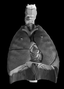
Trachea and Bronchi
Tracheal Structure
Trachea: Windpipe, a tube supported by C-shaped cartilages, conducting air to the bronchi.
Carina: The ridge at the base of the trachea where it divides into the right and left primary bronchi.
Bronchi: Branches of the trachea (primary, secondary, tertiary) that conduct air into the lungs.
Vocal Cords: True and false vocal cords are involved in sound production and airway protection.
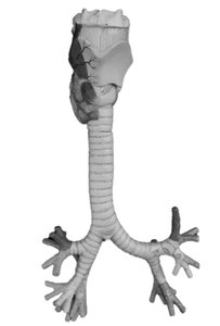
Lung Lobes and Fissures
Lobes of the Lungs
Right Lung: Three lobes – upper, middle, and lower.
Left Lung: Two lobes – upper and lower; contains the cardiac notch for the heart.
Fissures: Oblique and horizontal fissures separate the lobes.
Hilum: The entry/exit site for bronchi, blood vessels, and nerves.


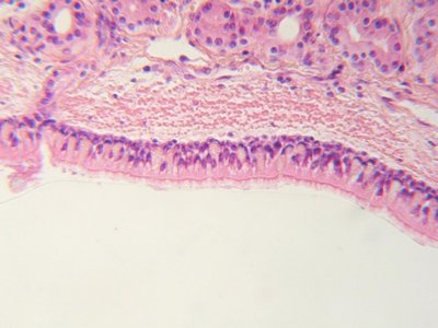
Upper Respiratory Tract Histology
Microscopic Structure
Mucosa: Lined with pseudostratified ciliated columnar epithelium, which helps trap and move particles out of the airway.
Submucosa: Areolar connective tissue containing glands.
Hyaline Cartilage: Provides structural support to the trachea.
Adventitia: Outermost connective tissue layer.
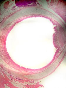
Specialized Cells
Cilia: Hair-like projections that move mucus and trapped particles upward.
Goblet Cells: Secrete mucus to trap debris.
Trachealis Muscle: Adjusts tracheal diameter.
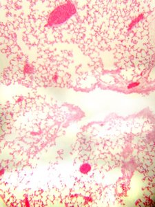
Lung Histology
Microscopic Anatomy
Bronchioles: Small airways lined with cuboidal epithelium and surrounded by smooth muscle.
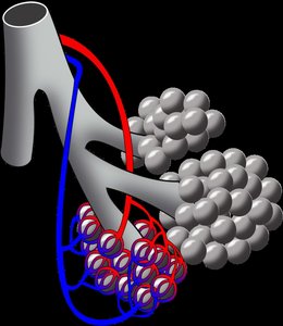
Alveoli: Tiny air sacs lined with simple squamous epithelium, the primary site of gas exchange.
Pulmonary Arteriole and Venule: Blood vessels involved in gas exchange.
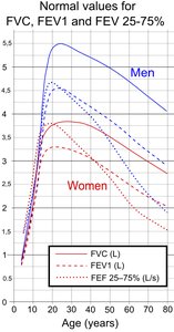
Lung Function Tests
Key Measurements
FVC (Forced Vital Capacity): The maximum amount of air a person can exhale after a maximal inhalation.
FEV1 (Forced Expiratory Volume in 1 second): The volume of air exhaled in the first second of a forced exhalation.
FEF25-75%: The average flow rate during the middle half of the FVC maneuver; reflects small airway function.
Equation:
This equation describes the formation of carbonic acid from carbon dioxide and water, which then dissociates into hydrogen and bicarbonate ions, important for acid-base balance in the blood.
Restrictive vs. Obstructive Disorders
Restrictive Disorders: Reduce lung inflation (e.g., pulmonary fibrosis); FVC is decreased.
Obstructive Disorders: Block airflow or increase airway resistance (e.g., COPD, asthma); FEV1 is decreased more than FVC.
Lung Volumes and Capacities
Tidal Volume: Normal breath volume.
Residual Volume: Air remaining after maximal exhalation.
Inspiratory/Expiratory Reserve Volume: Additional air that can be inhaled/exhaled after a normal breath.
Total Lung Capacity: Sum of all lung volumes.
Histology of Respiratory Diseases
Asthma
Bronchial Asthma: Characterized by broncho-constriction and increased mucus production, leading to narrowed airways and difficulty breathing.
Emphysema
Emphysema: A chronic obstructive pulmonary disease marked by the breakdown of alveolar walls, resulting in larger but fewer alveoli and reduced surface area for gas exchange.
Additional info: This guide integrates anatomical, histological, and functional aspects of the respiratory system, as well as clinical correlations with common respiratory diseases.
