 Back
BackChapter 25: The Urinary System – Structure, Function, and Physiology
Study Guide - Smart Notes
 Tailored notes based on your materials, expanded with key definitions, examples, and context.
Overview of the Urinary System
Main Functions of the Urinary System
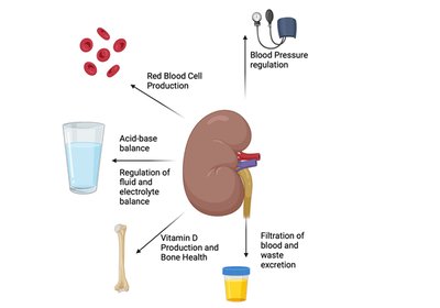
The urinary system is essential for maintaining homeostasis by regulating the composition and volume of blood, removing waste products, and balancing electrolytes and pH. The kidneys are the primary organs responsible for these functions.
Regulation of body water volume, blood osmolarity, and ion concentration: Kidneys filter excess water and ions, conserving or excreting them as needed to maintain osmotic balance.
Regulation of blood pressure: By controlling blood volume and releasing the hormone renin, kidneys influence systemic blood pressure.
Acid-base balance: Kidneys regulate blood pH by excreting or generating bicarbonate and hydrogen ions.
Excretion of metabolic waste, toxins, and drugs: The kidneys clear the blood of metabolic byproducts and foreign substances.
Production of erythropoietin (EPO): EPO stimulates red blood cell production in response to hypoxia.
Activation of vitamin D: Kidneys convert vitamin D to its active form, calcitriol, which is crucial for calcium absorption.
Gluconeogenesis: During prolonged fasting, kidneys can generate glucose from non-carbohydrate sources.
Anatomy of the Urinary System
Major Structures
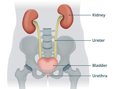
The urinary system consists of the kidneys, ureters, urinary bladder, and urethra. Each structure plays a specific role in urine formation, transport, storage, and elimination.
Kidneys: Filter blood and form urine.
Ureters: Paired tubes that transport urine from the kidneys to the bladder.
Urinary Bladder: Temporary storage reservoir for urine.
Urethra: Tube that carries urine from the bladder to the exterior of the body.
Gross Anatomy of the Kidneys
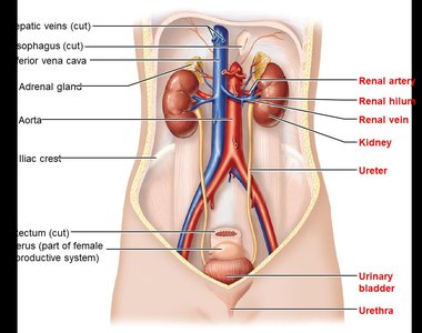
The kidneys are retroperitoneal organs located on the posterior abdominal wall, typically between the T12 and L3 vertebrae. The right kidney is slightly lower than the left due to the position of the liver. Each kidney is capped by an adrenal gland.
Lateral surface: Convex
Medial surface: Concave, containing the renal hilum (entry/exit for vessels, nerves, and ureter)
Internal Structure of the Kidney
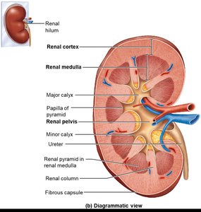
The kidney is divided into three main regions:
Renal Cortex: The outer, lighter-colored region where most nephrons are located.
Renal Medulla: The middle region, composed of renal pyramids separated by renal columns.
Renal Pelvis: The innermost region, a funnel-shaped structure that collects urine and channels it into the ureter.
Blood Supply of the Kidneys
The kidneys receive a rich blood supply, essential for filtration and homeostatic regulation. Blood flows through a series of arteries and capillaries, including the renal artery, segmental arteries, interlobar arteries, arcuate arteries, and cortical radiate arteries, before entering the glomerulus for filtration.
Nephrons: The Functional Units of the Kidney
Structure of the Nephron
Nephrons are the microscopic structural and functional units of the kidney, responsible for filtering blood and forming urine. Each nephron consists of a renal corpuscle and renal tubule.
Renal Corpuscle: Includes the glomerulus (a ball of fenestrated capillaries) and the glomerular (Bowman's) capsule, which collects the filtrate.
Renal Tubule: Composed of the proximal convoluted tubule (PCT), nephron loop (loop of Henle), and distal convoluted tubule (DCT).
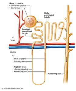
Types of Nephrons
Cortical Nephrons: Make up 85% of nephrons; located mostly in the cortex.
Juxtamedullary Nephrons: 15% of nephrons; have long loops that extend deep into the medulla, crucial for concentrating urine.
Nephron Capillary Beds
Peritubular Capillaries: Surround most of the renal tubule, involved in reabsorption and secretion.
Vasa Recta: Surround the loop of Henle in juxtamedullary nephrons, important for maintaining the medullary osmotic gradient.
Renal Physiology: Urine Formation
Major Renal Processes
Urine formation involves three main processes:
Filtration: Occurs at the glomerulus, producing a cell- and protein-free filtrate.
Reabsorption: Movement of substances from the filtrate back into the blood, primarily in the PCT.
Secretion: Additional substances are secreted into the tubule for excretion.
Filtration Membrane
The filtration membrane consists of three layers:
Fenestrated endothelium of glomerular capillaries: Allows passage of small solutes but not blood cells.
Basement membrane: Negatively charged, repels most proteins.
Podocytes: Specialized cells with filtration slits that further restrict passage of large molecules.
Filtration Pressures
Filtration is driven by hydrostatic pressure in the glomerular capillaries, opposed by capsular hydrostatic pressure and blood colloid osmotic pressure. The net filtration pressure determines the glomerular filtration rate (GFR).
Equation for Net Filtration Pressure (NFP):
Where:
= Hydrostatic pressure in glomerular capillaries
= Hydrostatic pressure in capsular space
= Osmotic pressure in glomerular capillaries
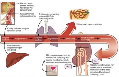
Regulation of Glomerular Filtration Rate (GFR)
GFR is regulated by intrinsic (renal autoregulation) and extrinsic (nervous and endocrine) mechanisms:
Myogenic response: Afferent arteriole constricts or dilates in response to blood pressure changes.
Tubuloglomerular feedback: Macula densa cells sense NaCl concentration and adjust afferent arteriole diameter.
Renin-angiotensin-aldosterone system (RAAS): Activated by low blood pressure, leading to increased sodium and water reabsorption.
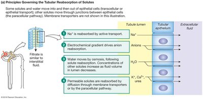
Tubular Reabsorption and Secretion
Mechanisms of Reabsorption
Most reabsorption occurs in the proximal convoluted tubule (PCT), where substances like glucose, amino acids, and ions are actively or passively transported back into the blood. Water reabsorption follows solute reabsorption via osmosis, aided by aquaporins.
Transcellular route: Through the epithelial cells.
Paracellular route: Between epithelial cells.
Hormonal Regulation
Antidiuretic hormone (ADH): Increases water reabsorption in the collecting duct by increasing aquaporin channels.
Aldosterone: Increases sodium (and thus water) reabsorption in the distal tubule and collecting duct.
Atrial natriuretic peptide (ANP): Inhibits sodium reabsorption, promoting natriuresis and diuresis.
Parathyroid hormone (PTH): Increases calcium reabsorption in the distal tubule.
Tubular Secretion
Secretion removes additional wastes and excess ions (e.g., H+, K+, NH4+, creatinine, drugs) from the blood into the tubular fluid, helping to regulate acid-base balance and eliminate toxins.
Concentration and Dilution of Urine
Countercurrent Mechanisms
The nephron loop and vasa recta establish and maintain a medullary osmotic gradient, allowing the kidneys to produce urine of varying concentration depending on hydration status.
Countercurrent multiplier: The loop of Henle creates the gradient by differential permeability to water and solutes.
Countercurrent exchanger: The vasa recta preserves the gradient by exchanging solutes and water with the interstitium.
Urea Recycling
Urea contributes to the medullary osmotic gradient by recycling between the collecting duct and nephron loop, enhancing the kidney's ability to concentrate urine.
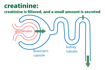
Evaluation of Renal Function
Renal Clearance
Renal clearance is the volume of plasma cleared of a substance per minute. It is used to estimate GFR and assess kidney function.
Renal Clearance Equation:
Where:
= clearance rate (ml/min)
= concentration of substance in urine
= urine flow rate (ml/min)
= concentration of substance in plasma
Physical and Chemical Properties of Urine
Composition: 95% water, 5% solutes (urea, uric acid, creatinine, ions)
Normal findings: No glucose, proteins, ketone bodies, hemoglobin, bile pigments, RBCs, or WBCs
Color: Clear to deep yellow (due to urobilin)
pH: Slightly acidic (pH ~6), but can range from 4.5 to 8.0
Specific gravity: 1.001–1.035 (higher than pure water)
Urinary Tract: Ureters, Bladder, and Urethra
Ureters
Ureters are muscular tubes that transport urine from the renal pelvis to the bladder via peristaltic contractions. They have three layers: mucosa (transitional epithelium), muscularis (smooth muscle), and adventitia (connective tissue).
Urinary Bladder
The bladder is a distensible, muscular sac that stores urine. It has rugae and transitional epithelium to allow expansion. The detrusor muscle contracts during urination. The trigone is a common site for urinary tract infections (UTIs).
Urethra
The urethra drains urine from the bladder to the exterior. It is shorter in females (3–4 cm) and longer in males (20 cm, with prostatic, membranous, and spongy regions). Two sphincters control urination: the internal (involuntary) and external (voluntary) urethral sphincters.
Micturition (Urination)
Micturition is the process of emptying the bladder. It involves contraction of the detrusor muscle and relaxation of both urethral sphincters, coordinated by the nervous system. Voluntary control develops around age 2–3.
Clinical Correlations
Glomerulonephritis: Inflammation of the glomerulus, leading to proteinuria and hematuria.
Nephrolithiasis/Ureterolithiasis: Kidney stones can obstruct urine flow and cause pain.
Urinary incontinence: Inability to control urination, often due to weakened pelvic muscles.
Urinary retention: Inability to expel urine, possibly due to anesthesia or prostate enlargement in males.
