 Back
BackChapter 26: The Reproductive System – Structure, Function, and Physiology
Study Guide - Smart Notes
 Tailored notes based on your materials, expanded with key definitions, examples, and context.
Chapter 26: The Reproductive System
Introduction to the Reproductive System
The reproductive system is essential for the production of offspring and the continuation of species. It consists of primary sex organs (gonads) and accessory reproductive organs. The gonads (testes in males, ovaries in females) produce gametes and secrete sex hormones, while accessory organs support gamete transport, fertilization, and development.
Gonads: Produce gametes (sperm and ova) and secrete hormones (testosterone, estrogens).
Accessory organs: Include ducts, glands, and external genitalia that facilitate reproduction.
Chromosome Sets and Ploidy
Chromosome number is described by the term "-ploid." Most human cells are diploid (2n), containing two sets of chromosomes, while gametes are haploid (1n), containing one set. This reduction in chromosome number is essential for maintaining genetic stability across generations.
Diploid (2n): Two sets of chromosomes (somatic cells).
Haploid (1n): One set of chromosomes (gametes: sperm and ova).
26.1 Overview of the Reproductive System and Meiosis
Comparison of Male and Female Reproductive Systems
Both systems share the same basic functions: gamete production, hormone secretion, and structures for fertilization and development. However, the organs and processes differ between sexes.
Male: Testes produce sperm and testosterone; ducts and glands support sperm maturation and delivery.
Female: Ovaries produce ova and estrogens; uterine tubes, uterus, and vagina support fertilization, gestation, and birth.
Mitosis vs. Meiosis
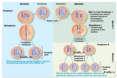
Mitosis and meiosis are two types of cell division. Mitosis produces genetically identical diploid cells for growth and repair, while meiosis produces genetically unique haploid gametes for sexual reproduction.
Mitosis: One division, two diploid daughter cells, identical to parent cell.
Meiosis: Two divisions, four haploid daughter cells, genetically unique due to crossing over and independent assortment.
Stages of Meiosis
Meiosis consists of two sequential divisions: Meiosis I (reduction division) and Meiosis II (equational division). Key events include homologous chromosome pairing, crossing over, and separation of chromatids.
Meiosis I: Homologous chromosomes separate, reducing chromosome number by half.
Meiosis II: Sister chromatids separate, producing four haploid cells.
26.2 Anatomy of the Male Reproductive System
Structure and Function
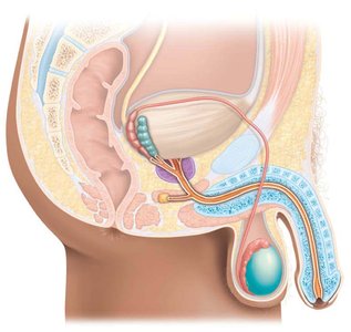
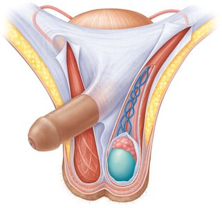
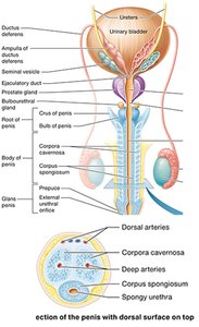
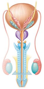
The male reproductive system includes the testes, ducts, accessory glands, and penis. The testes are the site of sperm production and hormone secretion.
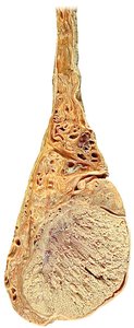
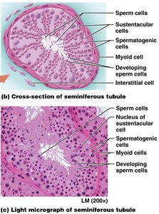
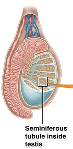
Testes: Contain seminiferous tubules (site of spermatogenesis) and interstitial cells (testosterone production).
Scrotum: Regulates testicular temperature for optimal sperm production.
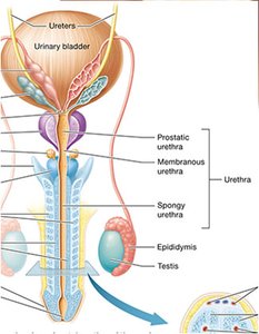
Ducts: Epididymis (sperm maturation), ductus deferens (sperm transport), ejaculatory ducts, and urethra (shared with urinary system).
Accessory glands: Seminal vesicles, prostate gland, and bulbourethral glands add secretions to semen.
Penis: Organ of copulation, contains erectile tissue and urethra.
Pathway of Sperm
Sperm are produced in the seminiferous tubules, mature in the epididymis, and travel through the ductus deferens, ejaculatory duct, and urethra during ejaculation.
Accessory Glands and Semen
Accessory glands secrete fluids that nourish and protect sperm, forming semen. Semen contains sperm, nutrients, and substances that enhance motility and survival.
Seminal vesicles: Fructose, prostaglandins, clotting proteins.
Prostate gland: Citrate, PSA, antimicrobial chemicals.
Bulbourethral glands: Mucus, alkaline secretions.
26.3 Physiology of the Male Reproductive System
Spermatogenesis
Spermatogenesis is the process of sperm production, involving mitosis, meiosis, and spermiogenesis. It occurs in the seminiferous tubules and is regulated by hormones.
Spermatogonia: Diploid stem cells that divide by mitosis.
Primary spermatocytes: Undergo meiosis I to form secondary spermatocytes (haploid).
Secondary spermatocytes: Undergo meiosis II to form spermatids (haploid).
Spermiogenesis: Maturation of spermatids into spermatozoa.
Hormonal Regulation
The hypothalamic-pituitary-gonadal axis controls spermatogenesis and testosterone production.
GnRH (Gonadotropin-releasing hormone): From hypothalamus, stimulates FSH and LH release from anterior pituitary.
FSH (Follicle-stimulating hormone): Stimulates spermatogenesis.
LH (Luteinizing hormone): Stimulates testosterone secretion from Leydig cells.
26.4 Anatomy of the Female Reproductive System
Structure and Function
The female reproductive system includes the ovaries, uterine tubes, uterus, vagina, vulva, and mammary glands. The ovaries produce ova and hormones, while other structures support fertilization, gestation, and childbirth.
Ovaries: Site of oogenesis and hormone production; anchored by ligaments.
Uterine tubes: Transport ova, site of fertilization.
Uterus: Site of implantation, fetal development, and labor; composed of endometrium, myometrium, and perimetrium.
Vagina: Passage for sperm, menstrual flow, and childbirth.
Vulva: External genitalia; homologous structures to male system.
Mammary glands: Modified sweat glands for milk production.
26.5 Physiology of the Female Reproductive System
Oogenesis
Oogenesis is the process of ovum formation, beginning before birth and continuing in cycles from puberty to menopause. It involves mitosis, meiosis, and follicular development.
Oogonia: Diploid stem cells; most degenerate before birth.
Primary oocytes: Arrested in prophase I until puberty.
Secondary oocytes: Complete meiosis I at ovulation; meiosis II completed only if fertilization occurs.
Ovarian and Uterine Cycles
The female reproductive cycle consists of the ovarian cycle (follicle development and ovulation) and the uterine cycle (endometrial changes). Hormonal regulation synchronizes these cycles.
Ovarian cycle: Follicular phase, ovulation, luteal phase.
Uterine cycle: Menstrual, proliferative, secretory phases.
Hormones: FSH, LH, estrogen, progesterone regulate cycle events.
Phases of the Menstrual Cycle
Phase | Ovarian Event | Uterine Event | Hormones |
|---|---|---|---|
Menstrual (Days 1-5) | Follicle development begins | Endometrium shed | Low estrogen/progesterone |
Pre-ovulatory (6-13) | Follicle matures | Endometrium proliferates | Rising estrogen |
Ovulation (~14) | Follicle ruptures, oocyte released | Endometrium thickens | LH surge, peak estrogen/FSH |
Post-ovulatory (15-28) | Corpus luteum forms | Endometrium prepares for implantation | High progesterone |
26.6 Methods of Birth Control
Birth control methods prevent pregnancy by blocking fertilization, inhibiting ovulation, or preventing implantation. Effectiveness varies with method and user compliance.
Barrier methods: Condoms, diaphragms.
Hormonal methods: Oral contraceptives, injections, vaginal rings.
Sterilization: Vasectomy, tubal ligation.
IUDs: Prevent implantation.
Chemical methods: Spermicides.
Behavioral methods: Rhythm, sympto-thermal methods.
26.7 Sexually Transmitted Infections (STIs)
STIs are caused by bacteria, parasites, or viruses and can disrupt reproductive health. Common examples include chlamydia, gonorrhea, syphilis, trichomoniasis (bacterial/parasitic), and HPV, genital herpes (viral).
Bacterial/parasitic STIs: Cause inflammation, discharge, tissue damage.
Viral STIs: Invade host cells, may cause chronic infection or cancer.
Comparison of Spermatogenesis and Oogenesis
Process | Timing | Number of Gametes | Duration |
|---|---|---|---|
Spermatogenesis | Begins at puberty | Millions per day | Continuous through life |
Oogenesis | Begins before birth | 1 per month | Stops at menopause |
Additional info: The reproductive system is closely regulated by hormonal feedback loops, and disruptions can affect fertility and overall health. Understanding the anatomy and physiology of both male and female systems is essential for clinical practice and health education.
