 Back
BackChapter 3: Cells – The Living Units (Mini-Textbook Study Notes)
Study Guide - Smart Notes
 Tailored notes based on your materials, expanded with key definitions, examples, and context.
Cells: The Living Units
Why This Matters
Understanding the structure and function of cells is fundamental to human anatomy and physiology. The permeability of the plasma membrane is crucial for cellular health and can influence medical treatments.
Cell Theory and Cell Diversity
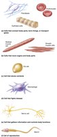
Cells are the basic structural and functional units of all living organisms. The human body contains trillions of cells, with over 250 distinct types differing in size, shape, and internal components, which determine their specialized functions.
Cell Theory: All living things are composed of cells; cells are the smallest unit of life; new cells arise only from pre-existing cells.
Cell Diversity: Variations in cell morphology and organelles lead to functional specialization.
Examples: Muscle cells contract, nerve cells transmit signals, epithelial cells cover surfaces.
Generalized Cell Structure
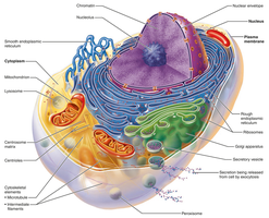
Despite diversity, all human cells share three fundamental components:
Plasma Membrane: Flexible boundary separating internal and external environments.
Cytoplasm: Intracellular fluid containing organelles.
Nucleus: Control center housing DNA.
Extracellular Materials
Substances outside cells include:
Extracellular fluids: Interstitial fluid, blood plasma, cerebrospinal fluid.
Cellular secretions: Saliva, mucus, gastric fluids.
Extracellular matrix: Structural support and cell adhesion.
Plasma Membrane Structure and Function
Fluid Mosaic Model
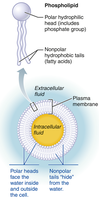
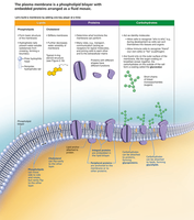
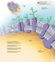
The plasma membrane is a dynamic, double layer of phospholipids with embedded proteins and surface carbohydrates. It acts as a selective barrier and facilitates cell communication.
Phospholipid Bilayer: Hydrophilic heads face water; hydrophobic tails face inward.
Cholesterol: Adds membrane stiffness.
Proteins: Integral and peripheral, each with specialized functions.
Glycocalyx: Surface sugars for cell recognition.
Membrane Proteins
Membrane proteins are essential for cell communication, transport, enzymatic activity, and structural support.
Integral Proteins: Span the membrane; function as channels, carriers, receptors, and enzymes.
Peripheral Proteins: Loosely attached; function in cell shape, division, and connections.
Functions of Membrane Proteins
Transport: Control entry/exit of substances. Channels and pumps facilitate movement.
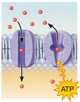
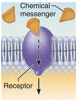
Receptors: Receive signals and initiate cellular responses (signal transduction).
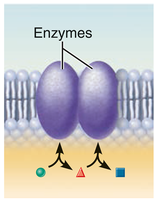
Enzymatic Activity: Catalyze reactions at the membrane.
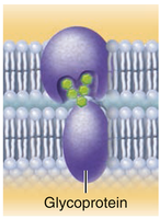
Cell-Cell Recognition: Glycoproteins serve as identification tags.
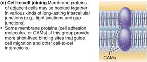
Cell-to-Cell Joining: Form junctions for tissue integrity and communication.
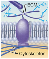
Attachment: Anchor cytoskeleton and extracellular matrix for structural stability.
Glycocalyx
The glycocalyx is a carbohydrate-rich area on the cell surface, functioning as a biological marker for cell recognition and immune response.
Glycolipids and Glycoproteins: Contribute to unique cell identity.
Clinical Note: Rapid changes in cancer cell glycocalyx can evade immune detection.
Intercellular Junctions
Types of Cell Junctions
Cell junctions allow cells to adhere and communicate, maintaining tissue integrity.
Tight Junctions: Impermeable barriers preventing passage between cells.
Desmosomes: Anchoring junctions providing mechanical strength.
Gap Junctions: Channels for direct communication and transfer of ions/molecules.
Membrane Transport
Passive Transport
Passive transport moves substances across the membrane without energy input, relying on diffusion down concentration gradients.
Simple Diffusion: Direct movement of lipid-soluble or small molecules.
Facilitated Diffusion: Assisted by carrier or channel proteins for larger or water-soluble molecules.
Osmosis: Movement of water through the membrane or aquaporins.
Osmolarity and Tonicity
Osmolarity measures solute concentration; tonicity describes a solution's effect on cell volume.
Isotonic: No net water movement; cell volume unchanged.
Hypertonic: Water leaves cell; cell shrinks (crenation).
Hypotonic: Water enters cell; cell swells (lysis).
Active Transport
Active transport requires ATP to move substances against their concentration gradient.
Primary Active Transport: Direct use of ATP (e.g., sodium-potassium pump).
Secondary Active Transport: Indirect use of ATP via ion gradients (symporters and antiporters).
Vesicular Transport
Vesicular transport moves large substances or volumes via vesicles, requiring energy.
Endocytosis: Intake of substances (phagocytosis, pinocytosis, receptor-mediated).
Exocytosis: Ejection of substances from the cell.
Membrane Potential and Cell Signaling
Resting Membrane Potential (RMP)
RMP is the voltage across the cell membrane, primarily established by potassium ion gradients. It is essential for nerve and muscle function.
Electrochemical Gradients: Maintained by sodium-potassium pumps.
Gated Channels: Opening alters membrane potential, activating cells.
Cell Adhesion Molecules (CAMs) and Membrane Receptors
CAMs anchor cells and facilitate movement, while membrane receptors mediate cell signaling and responses to external stimuli.
Contact Signaling: Recognition via surface receptors.
Chemical Signaling: Ligand-receptor interactions trigger cellular changes.
G Protein–Coupled Receptors: Indirectly activate intracellular messengers.
Cell Life Cycle and Aging
Autophagy, Proteasomes, and Apoptosis
Cells dispose of damaged organelles and proteins via autophagy and proteasomes. Apoptosis is programmed cell death, essential for removing unneeded or damaged cells.
Autophagy: Self-eating process for organelle turnover.
Proteasomes: Degrade ubiquitin-tagged proteins.
Apoptosis: Controlled cell death via caspase activation.
Cell Differentiation and Aging
Cells differentiate during development, guided by chemical signals. Aging theories include wear and tear, mitochondrial dysfunction, immune decline, and genetic programming. Telomeres limit cell division; telomerase can extend telomeres, contributing to cancer cell immortality.
Hyperplasia: Increased cell numbers for growth.
Atrophy: Decreased cell size from loss of stimulation.
Progeria: Rare disease mimicking aging due to nuclear instability.
Additional info: These notes expand on brief textbook points to provide a comprehensive, exam-ready overview of cell structure, function, and physiology, suitable for ANP college students.
