 Back
BackChapter 3: Cells – The Living Units (BSC 2085 Human Anatomy and Physiology I)
Study Guide - Smart Notes
 Tailored notes based on your materials, expanded with key definitions, examples, and context.
Cells: The Living Units
Overview of Cells
Cells are the basic structural and functional units of all living organisms. Human cells share common features but also exhibit diversity in structure and function, reflecting their specialized roles in the body.
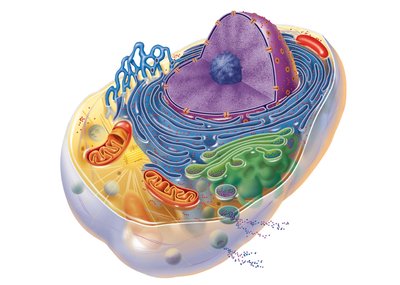
Generalized Eukaryotic Cell: All human cells have three main parts: the plasma membrane, cytoplasm, and nucleus.
Cell Diversity: Cells vary in shape, size, and function (e.g., fibroblasts, erythrocytes, nerve cells, muscle cells, fat cells, sperm).
The Plasma Membrane
Structure and Function
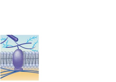
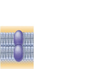
The plasma membrane is a flexible, dynamic boundary that separates the intracellular fluid (ICF) from the extracellular fluid (ECF). It is composed of a bimolecular layer of lipids and proteins, forming a fluid mosaic model.
Phospholipid Bilayer: 75% phospholipids (hydrophilic heads, hydrophobic tails), 5% glycolipids, 20% cholesterol (stabilizes membrane).
Integral Proteins: Firmly embedded, often span the membrane (transmembrane); function as transporters, receptors, or enzymes.
Peripheral Proteins: Loosely attached to integral proteins; function as enzymes, motor proteins, or in cell-to-cell connections.
Glycocalyx: Carbohydrate-rich area on the cell surface important for recognition and interaction.
Functions of Membrane Proteins
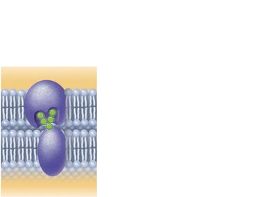
Transport: Channels and carriers move substances across the membrane.
Receptors for Signal Transduction: Bind chemical messengers and initiate cellular responses.
Attachment to Cytoskeleton and ECM: Maintain cell shape and stabilize location.
Enzymatic Activity: Catalyze metabolic reactions.
Intercellular Joining: Form cell junctions for tissue integrity.
Cell-Cell Recognition: Allow cells to identify each other.
Membrane Junctions
Specialized structures connect adjacent cells:
Tight Junctions: Prevent passage of molecules between cells.
Desmosomes: Anchor cells together, providing mechanical stability.
Gap Junctions: Allow communication and passage of ions/small molecules between cells.
Membrane Transport
Types of Membrane Transport
The plasma membrane is selectively permeable, allowing some substances to pass while restricting others. Transport occurs via passive or active processes.
Passive Processes: No energy required; substances move down their concentration gradient.
Active Processes: Require ATP; substances move against their concentration gradient or involve bulk transport.
Passive Transport
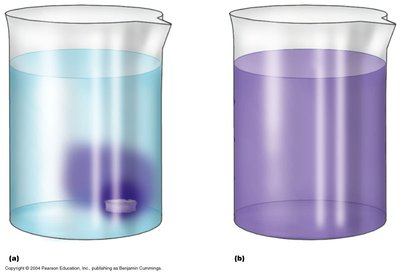
Simple Diffusion: Movement of lipid-soluble molecules directly through the lipid bilayer.
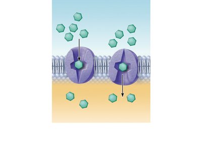
Facilitated Diffusion: Movement of lipid-insoluble molecules via protein carriers or channels.
Osmosis: Diffusion of water through a selectively permeable membrane.
Osmosis and Tonicity
Osmolarity: Total concentration of solute particles in a solution.
Isotonic Solution: Same solute concentration as the cell; no net water movement.
Hypertonic Solution: Higher solute concentration than the cell; cell shrinks (crenates).
Hypotonic Solution: Lower solute concentration than the cell; cell swells and may burst (lyse).
Active Transport
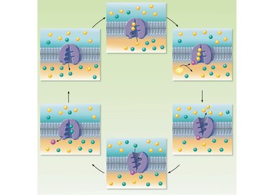
Primary Active Transport: Direct use of ATP to move substances (e.g., Na+-K+ pump).
Secondary Active Transport: Indirect use of ATP; movement of one substance drives movement of another.
Vesicular Transport
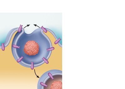
Endocytosis: Cell engulfs material via vesicles (phagocytosis for solids, pinocytosis for fluids).
Exocytosis: Vesicles fuse with the plasma membrane to release contents outside the cell.
The Cytoplasm and Organelles
Cytoplasm
The cytoplasm is the cellular material between the plasma membrane and the nucleus. It consists of cytosol (fluid), organelles (metabolic machinery), and inclusions (stored nutrients, pigments).
Membranous Organelles
Mitochondria: Site of ATP production via aerobic respiration; contain their own DNA and RNA.
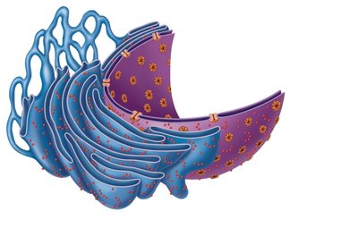
Endoplasmic Reticulum (ER): Network of membranes; rough ER (with ribosomes) synthesizes proteins, smooth ER synthesizes lipids and detoxifies chemicals.
Golgi Apparatus: Modifies, sorts, and packages proteins and lipids for secretion or delivery to other organelles.
Lysosomes: Contain digestive enzymes; break down waste and cellular debris.
Peroxisomes: Contain oxidases and catalases; detoxify harmful substances.
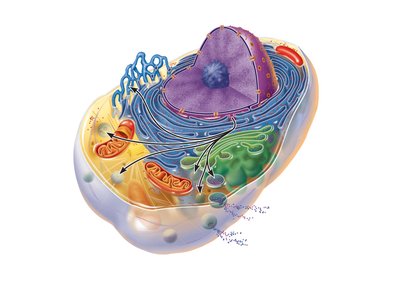
Nonmembranous Organelles
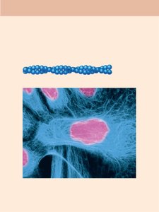
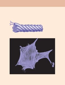
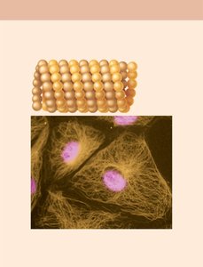
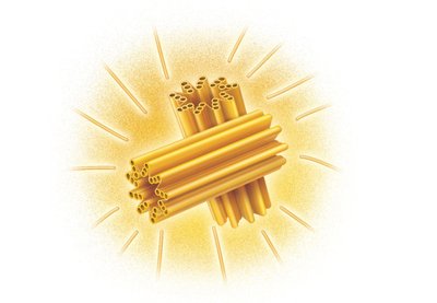
Cytoskeleton: Network of protein filaments (microfilaments, intermediate filaments, microtubules) providing structural support and movement.
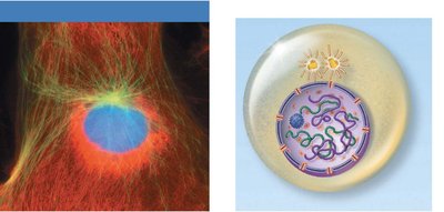
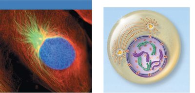
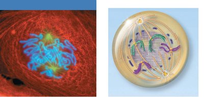
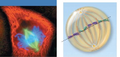
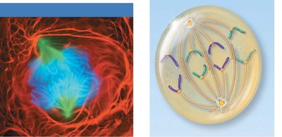
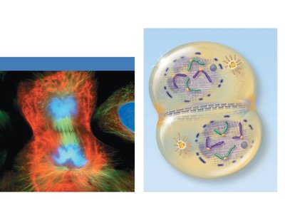
Centrosome and Centrioles: Organize microtubules and form the mitotic spindle during cell division.
Ribosomes: Sites of protein synthesis; free in cytosol or bound to rough ER.
Cellular Extensions
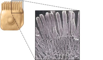
Microvilli: Increase surface area for absorption; core of actin filaments.
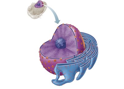
The Nucleus
Structure and Function
The nucleus is the control center of the cell, containing the genetic material (DNA) and directing cellular activities.
Nuclear Envelope: Double membrane with nuclear pores for transport.
Nucleolus: Site of rRNA synthesis and ribosome assembly.
Chromatin: DNA and associated proteins; condenses to form chromosomes during cell division.
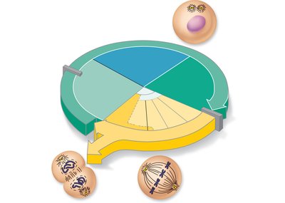
The Cell Life Cycle (Mitosis)
Overview
The cell cycle consists of interphase (growth and DNA replication) and the mitotic phase (division of the nucleus and cytoplasm).
Interphase: G1 (growth), S (DNA synthesis), G2 (preparation for division).
Mitosis: Prophase, Metaphase, Anaphase, Telophase.
Cytokinesis: Division of the cytoplasm, forming two daughter cells.

DNA Structure and Function
DNA Organization
DNA is a double helix composed of nucleotides (deoxyribose sugar, phosphate group, nitrogenous base). It is organized into chromatin and chromosomes.
Base Pairing: Adenine pairs with thymine, guanine pairs with cytosine.
Nucleosome: DNA wrapped around histone proteins.
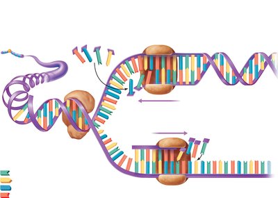
DNA Replication
During the S phase of interphase, DNA is replicated to ensure each daughter cell receives an identical set of genetic instructions.
Enzymes: Helicase unwinds DNA; DNA polymerase synthesizes new strands.
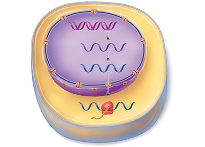
Protein Synthesis
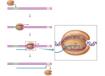
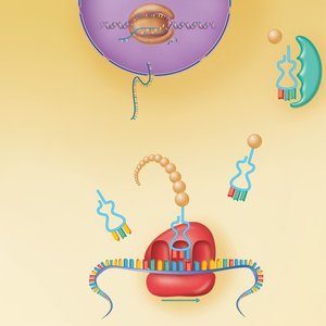
Transcription and Translation
Transcription: DNA is used as a template to synthesize messenger RNA (mRNA) in the nucleus.
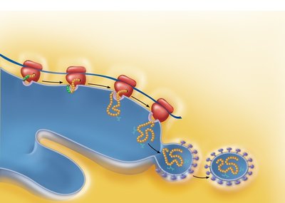
Translation: mRNA is decoded by ribosomes in the cytoplasm to assemble amino acids into a polypeptide chain.
Types of RNA: mRNA (carries code), rRNA (structural component of ribosomes), tRNA (brings amino acids to ribosome).
Summary Table: Key Cell Structures and Functions
Structure | Main Function |
|---|---|
Plasma Membrane | Selective barrier, cell communication |
Nucleus | Genetic control center |
Mitochondria | ATP production |
Ribosomes | Protein synthesis |
Rough ER | Protein modification and transport |
Smooth ER | Lipid synthesis, detoxification |
Golgi Apparatus | Protein and lipid packaging |
Lysosomes | Digestion of macromolecules |
Peroxisomes | Detoxification |
Cytoskeleton | Structural support, movement |
