 Back
BackChapter 4: Histology – The Study of Tissues
Study Guide - Smart Notes
 Tailored notes based on your materials, expanded with key definitions, examples, and context.
Histology: The Study of Tissues
Introduction to Tissues
Histology is the study of the normal structures of tissues, which are groups of structurally and functionally related cells and their external environment. All tissues share two basic components: a discrete population of cells and the extracellular matrix (ECM), which surrounds the cells.
Discrete population of cells: Cells are related in structure and function.
Extracellular matrix (ECM): The material surrounding cells, providing structural and functional support.
Types of Tissues
There are four primary tissue types, each defined by the kind and number of cells, the amount and composition of ECM, and their specific functions:
Epithelial tissues: Tightly packed sheets of cells with no visible ECM; cover and line all body surfaces and cavities. Specialized epithelia form glands that manufacture secretions such as sweat, saliva, or hormones.
Connective tissues: Connect all other tissues to one another; ECM is a prominent feature. Cells are scattered throughout and serve to bind, support, protect, and allow for transportation of substances.
Muscle tissues: Capable of generating force by contracting; little ECM between cells.
Nervous tissues: Capable of generating, sending, and receiving messages; cells that support this activity are also within a unique ECM.
The Extracellular Matrix (ECM)
Components and Functions
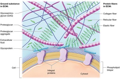
The ECM is a collection of substances in liquid, thick gel, or solid form that surround the cells of tissue. It consists of two main components: ground substance and protein fibers.
Ground substance: Extracellular fluid (ECF) with water, nutrients, ions, and three families of macromolecules: glycosaminoglycans (GAGs), proteoglycans, and cell-adhesion molecules (CAMs).
Protein fibers: Collagen fibers, elastic fibers, and reticular fibers, each providing different structural properties.
ECM functions include providing tissue strength, directing cells to proper positions, holding cells in place, and regulating development, mitotic activity, and survival.

Ground Substance
Glycosaminoglycans (GAGs): Examples include chondroitin sulfate and hyaluronic acid. Their negative charges attract positively charged ions, creating concentration gradients and drawing water out of cells and blood vessels by osmosis, helping ECM resist compression.
Proteoglycans: GAGs bound to a protein core, forming large aggregates that make ECM firmer and more resistant to compression. They also form barriers to diffusion, protecting underlying tissue.
Cell-adhesion molecules (CAMs): Glycoproteins that adhere cells to each other and to the ECM, maintaining tissue architecture.
Protein Fibers
Collagen fibers: Provide resistance to tension and pressure.
Elastic fibers: Composed of elastin, allow extensibility and elasticity.
Reticular fibers: Thin, short collagen fibers forming meshworks that support cells and ground substance.
Cell Junctions
Types of Cell Junctions
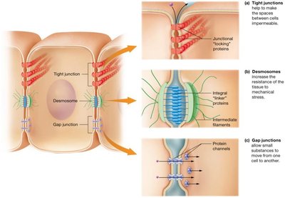
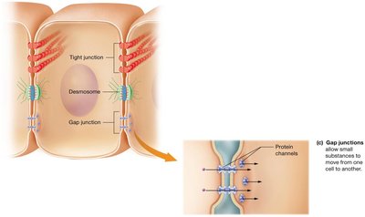
Cell junctions are ways cells bind to one another, with neighboring cell plasma membranes linked by integral proteins. There are three major types:
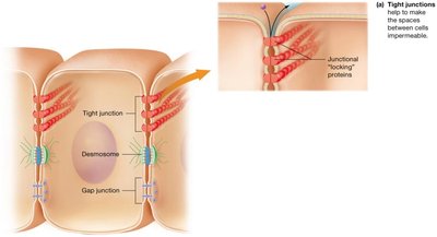
Tight junctions: Hold cells closely together, making the space between them impermeable to macromolecules.
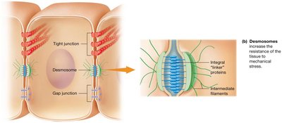
Desmosomes: Allow materials in extracellular fluid to pass through space between cells, increasing tissue strength.
Gap junctions: Small pores formed by protein channels, allowing small substances to flow freely between cells.
Tight Junctions
Tight junctions (occluding junctions) lock integral proteins of adjacent cell membranes together, forming a seal around the apical perimeter of the cell. This seal may not be complete, allowing for some leakage in certain tissues.
Desmosomes
Desmosomes link integral proteins and allow for passage of materials in extracellular fluid. They increase tissue strength by holding cells together and are attached to intermediate filaments for structural reinforcement.
Gap Junctions
Gap junctions are small pores formed by protein channels between adjacent cells, allowing small substances to flow freely between cytoplasms. They are important in tissues that communicate with electrical signals, such as cardiac muscle cells.
Epithelial Tissues
Functions of Epithelial Tissues
Epithelial tissues cover every internal and external body surface, acting as barriers between the body and the external environment. They line organs and fluid-filled cavities and serve several functions:
Protection: Shield underlying tissues from mechanical and thermal injury.
Immune defenses: Form physical barriers and prevent invasion by microorganisms.
Secretion: Form glands that produce substances like hormones and oils.
Transport: Selectively permeable membranes allow substances to cross barriers by passive or active transport.
Sensation: Rich nerve supply allows detection of changes in internal and external environments.
Components and Classification of Epithelia
Epithelial tissues consist of tightly packed cells linked by tight junctions and desmosomes, making sheets of cells fairly impermeable and resistant to physical stresses. They are avascular and rely on diffusion for nutrients.
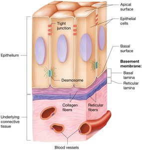
The ECM is found beneath cells in a thin basement membrane, composed of basal lamina (collagen fibers and ground substance) and reticular lamina (reticular fibers and ground substance). These layers anchor epithelial tissue to underlying connective tissue.
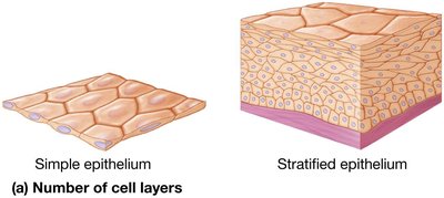
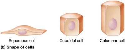
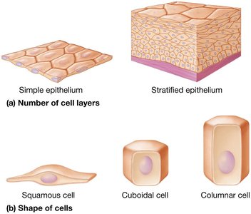
Classification Criteria
Number of cell layers: Simple epithelia (single layer), stratified epithelia (multiple layers).
Shape of cells: Squamous (flattened), cuboidal (short), columnar (tall and elongated).
Covering and Lining Epithelia
Simple Epithelia
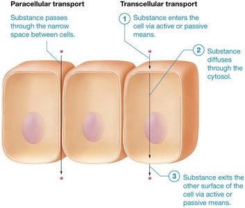
Simple epithelia are one cell-layer thick and adapted for transportation of substances between tissues. Some have microvilli for increased surface area, while others have cilia to move substances through hollow organs.
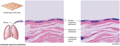
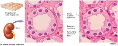
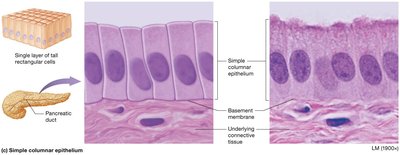
Transport Across Simple Epithelia
Substances can cross simple epithelia by paracellular transport (through narrow spaces between cells) or transcellular transport (through the cell itself).
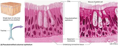
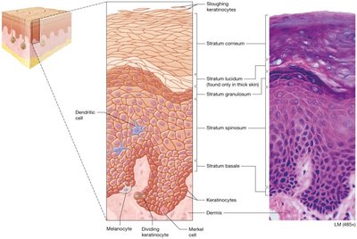
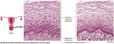
Stratified Epithelia
Stratified epithelia consist of multiple cell layers and are adapted for protection against mechanical stress.
Keratinized stratified squamous epithelium: Found in the epidermis, provides protection.
Nonkeratinized stratified squamous epithelium: Found in moist areas such as the vagina.
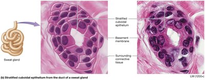
Stratified cuboidal and columnar epithelium: Found in ducts of glands.
Transitional epithelium: Specialized for stretching, found in the urinary bladder.
Glandular Epithelia
Types of Glands
Glands are structures of epithelial origin that synthesize and secrete products. They are classified by shape and by how products are released:
Endocrine glands: Secrete products (usually hormones) directly into the bloodstream, allowing systemic effects.
Exocrine glands: Release products onto apical surfaces or lining hollow organs via ducts, having local effects.
Unicellular and Multicellular Exocrine Glands
Goblet cells are the most common unicellular exocrine gland, secreting mucus in the digestive and respiratory tracts. Most exocrine glands are multicellular, classified by duct structure (simple or compound) and shape of secretory clusters (tubular, acinar, or tubuloacinar).
Type | Structure | Function |
|---|---|---|
Simple gland | Unbranched duct | Local secretion |
Compound gland | Branched duct | Local secretion |
Tubular | Long, straight/coiled | Secretion |
Acinar | Spherical | Secretion |
Tubuloacinar | Both tubular and acinar | Secretion |
*Additional info: Table inferred for clarity based on gland classification criteria.*
Connective Tissues
Types and Functions
Connective tissues are divided into connective tissue proper and specialized connective tissue. Their functions include connecting and binding, support, protection, and transport.
Connective tissue proper: Widely distributed, connects tissues and organs, and forms internal architecture.
Specialized connective tissue: Includes cartilage, bone, and blood, each with specific functions.
Cells of Connective Tissue Proper
Fibroblasts: Synthesize ECM and collagen.
Adipocytes: Store fat.
Mast cells: Involved in immune response.
Phagocytes: Engulf pathogens and debris.
Types of Connective Tissue Proper
Loose connective tissue: Areolar tissue, supports and binds other tissues.
Dense connective tissue: Irregular, regular, and elastic types, provide strength and flexibility.
Reticular tissue: Forms supportive networks in organs.
Adipose tissue: Stores energy and provides insulation.
Specialized Connective Tissues
Cartilage
Cartilage is a tough, flexible tissue found in joints, ear, nose, and respiratory tract. It absorbs shock and resists tension, compression, and shearing forces. It is populated by chondroblasts (immature cells) and chondrocytes (mature cells).
Bone Tissue
Bone tissue supports the body, protects organs, provides muscle attachments, stores calcium, and houses bone marrow. It consists of osteoblasts (bone-builders), osteocytes (mature cells), and osteoclasts (bone destroyers).
Blood
Blood is a unique connective tissue with a liquid ECM (plasma), consisting of water, dissolved solutes, and proteins. It serves as the main transport medium in the body.
Muscle Tissues
Types and Functions
Muscle tissues are specialized for contraction and include three types:
Skeletal muscle: Striated, voluntary movement.
Cardiac muscle: Striated, involuntary, found in the heart.
Smooth muscle: Non-striated, involuntary, found in walls of hollow organs.
Nervous Tissues
Components and Functions
Nervous tissue makes up the brain, spinal cord, and nerves. It consists of neurons (capable of sending and receiving messages) and neuroglial cells (support neuron activities). The ECM is unique, with specialized proteoglycans and few protein fibers.
Membranes
Types and Functions
Membranes are thin sheets of one or more tissues that line body surfaces or cavities. Most consist of a superficial epithelial layer resting on a connective tissue layer and sometimes contain smooth muscle. Functions include anchoring organs, serving as barriers, immunity, and secretion.
True membranes: Serous and synovial membranes.
Membrane-like structures: Mucous and cutaneous membranes.
Tissue Repair
Regeneration and Fibrosis
Tissue repair depends on the ability of resident cells to undergo mitosis. Epithelial tissues typically regenerate, while most connective tissues heal by regeneration except cartilage, which heals by fibrosis. Smooth muscle usually regenerates, but cardiac and skeletal muscle generally heal by fibrosis. Nervous tissue generally undergoes fibrosis, with neuroglial cells replacing dead neurons with scar tissue.
Factors Affecting Tissue Repair
Nutrition: Adequate supply of amino acids and vitamin C is required for collagen production.
Blood supply: Necessary for delivering oxygen, nutrients, and immune cells to the injured region.
