 Back
BackChapter 4: Histology – The Study of Tissues
Study Guide - Smart Notes
 Tailored notes based on your materials, expanded with key definitions, examples, and context.
Histology: The Study of Tissues
Definition and Importance
Histology is the study of the normal structures of tissues. All tissues share two basic components: a population of cells and the extracellular matrix (ECM), which is the material surrounding the cells. Tissues are organized into four primary types, each with distinct functions and structural characteristics.
Types of Tissues
Overview of the Four Primary Tissue Types
Epithelial tissues (epithelia): Tightly packed sheets of cells with no visible ECM; cover and line all body surfaces and cavities. Specialized epithelia form glands for secretion.
Connective tissues: Connect all other tissues to one another; ECM is a prominent feature. Functions include binding, support, protection, and transportation of substances.
Muscle tissues: Capable of generating force by contracting; little ECM between cells.
Nervous tissues: Capable of generating, sending, and receiving messages; unique ECM supports activity.
Organs are composed of multiple tissue types working together to perform specific functions (e.g., the heart contains muscle, epithelial, and connective tissues).
Extracellular Matrix (ECM)
Definition and Components
The extracellular matrix (ECM) is composed of substances surrounding the cells in a tissue. It provides strength, directs cell positioning, and holds cells in place. The ECM consists of two main components: ground substance and protein fibers.
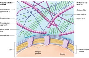
Ground Substance
Gel-like consistency containing extracellular fluid (ECF), water, nutrients, ions, and three families of macromolecules:
Glycosaminoglycans (GAGs): Long polysaccharide chains (e.g., chondroitin sulfate, hyaluronic acid). Their negative charge attracts positive ions, creating an osmotic gradient that draws water into the ECM, producing a gel-like matrix that resists compression.
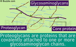
Proteoglycans: GAGs bound to a protein core, forming large aggregates. These make the ECM firmer and more resistant to compression, and act as barriers to diffusion, protecting tissues from microorganisms.
Cell-adhesion molecules (CAMs): Glycoproteins that adhere cells to each other and to ECM components, maintaining tissue structure.
Protein Fibers
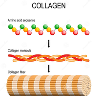
Collagen fibers: Comprise 20–25% of all proteins in the body; provide resistance to tension and pressure.
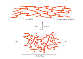
Elastic fibers: Composed of elastin; can stretch and return to original length (elasticity).
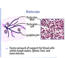
Reticular fibers: Thin, short collagen fibers forming a meshwork that supports cells and ground substance, especially in organs like the spleen and lymph nodes.
Cell Junctions
Types of Cell Junctions
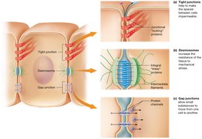
Cell junctions are specialized structures that bind cells together. There are three major types:
Tight junctions: Hold cells closely together, making the space between them impermeable to macromolecules. Example: blood vessel endothelium.
Desmosomes: Link integral proteins, allowing materials to pass between cells and distributing mechanical stress. Example: skin epithelium.
Gap junctions: Small pores formed by protein channels, allowing small substances to flow between cells. Example: cardiac muscle cells.
Epithelial Tissues
Functions and Characteristics
Epithelial tissues cover every internal and external body surface, acting as barriers and lining organs and cavities. Their functions include:
Protection: Shield underlying tissues from injury.
Immune defense: Form barriers against microorganisms; contain immune cells.
Secretion: Form glands that produce hormones, oils, etc.
Transport: Selectively permeable membranes for substance movement.
Sensation: Rich nerve supply for detecting environmental changes.
Classification of Epithelia
Number of layers: Simple (one layer) or stratified (multiple layers).
Cell shape: Squamous (flat), cuboidal (cube-shaped), columnar (tall and elongated).
Glandular Epithelia
Types of Glands
Endocrine glands: Secrete hormones directly into the bloodstream; systemic effects.
Exocrine glands: Release products onto body surfaces or into ducts; local effects. Can be unicellular (e.g., goblet cells) or multicellular (with various duct and secretory cell arrangements).
Modes of Secretion
Merocrine (eccrine): Products released by exocytosis (e.g., sweat glands).
Holocrine: Products released when cells rupture and die (e.g., sebaceous glands).
Apocrine: Portions of cytoplasm are pinched off with the product (e.g., mammary glands).
Connective Tissues
Classification and Functions
Connective tissues are divided into connective tissue proper and specialized connective tissues. Their functions include connecting and binding, support, protection, and transport.
Connective Tissue Proper
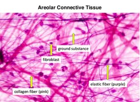
Loose connective tissue (areolar): Contains all three fiber types, fibroblasts, and ground substance; supports epithelia and houses immune cells.
Dense connective tissue: High density of protein fibers; includes dense irregular, dense regular, and dense elastic types.
Reticular tissue: Network of reticular fibers supporting small structures and organs.
Adipose tissue: Stores fat, insulates, and provides shock absorption.
Specialized Connective Tissues
Cartilage: Tough, flexible; three types (hyaline, fibrocartilage, elastic).
Bone: Rigid, supports body, stores calcium, houses marrow.
Blood: Fluid ECM (plasma); transports substances and cells.
Muscle Tissues
Types and Functions
Skeletal muscle: Voluntary, striated, multinucleate; moves the skeleton.
Cardiac muscle: Involuntary, striated, branched; found in the heart, contains intercalated discs for synchronized contraction.
Smooth muscle: Involuntary, non-striated; found in walls of hollow organs.
Nervous Tissues
Structure and Function
Neurons: Excitable cells that send and receive messages; consist of a cell body, axon, and dendrites.
Neuroglial cells: Support, protect, and nourish neurons; capable of mitosis.
Membranes
Types of Membranes
Serous membranes: Line body cavities not open to the exterior; secrete serous fluid to reduce friction.
Synovial membranes: Line joint cavities; secrete synovial fluid for lubrication.
Mucous membranes: Line passages open to the exterior; secrete mucus for protection.
Cutaneous membrane: The skin; protects underlying tissues.
Tissue Repair
Regeneration and Fibrosis
Regeneration: Replacement of dead or damaged cells with the same cell type; restores normal function (e.g., epithelial tissues, most connective tissues).
Fibrosis: Replacement with dense irregular connective tissue (scar tissue); some loss of function (e.g., cardiac muscle, nervous tissue).
Factors affecting tissue repair include the ability of cells to undergo mitosis, nutrition (especially protein and vitamin C), and blood supply.
