 Back
Chapter 4: Histology – The Study of Tissues
Study Guide - Smart Notes
 Tailored notes based on your materials, expanded with key definitions, examples, and context.
Histology: The Study of Tissues
Introduction to Tissues
Histology is the study of the normal structure of tissues, which are groups of structurally and functionally related cells and their external environment that together perform common functions. All tissues share two basic components: a discrete population of cells related in structure and function, and the surrounding material called the extracellular matrix (ECM).
Tissue: Group of related cells and their ECM performing a common function.
Histology: Study of tissue structure.
Extracellular Matrix (ECM): Material surrounding cells, providing support, regulating development, and holding cells in place.
Types of Tissues
Primary Tissue Types
There are four primary tissue types in the human body, each with distinct structures and functions:
Epithelial Tissues: Sheets of tightly packed cells with little ECM; cover and line body surfaces and cavities, and form parts of glands.
Connective Tissues: Connect all other tissues together; cells are scattered through the ECM; bind, support, protect, and allow transport of substances.
Muscle Tissues: Cells contract and generate force; little ECM.
Nervous Tissues: Neurons generate, send, and receive messages; includes supporting cells with a unique ECM.
Extracellular Matrix (ECM)
Components and Functions
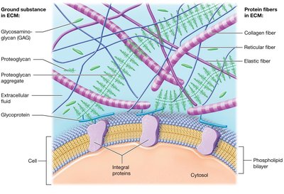
The ECM is composed of substances surrounding the cells in a tissue, providing strength, directing cell placement, regulating cell activity, and holding cells in position. It consists of two main components: ground substance and protein fibers.
Ground Substance: Gel-like substance with extracellular fluid, water, ions, nutrients, and macromolecules (glycosaminoglycans, proteoglycans, glycoproteins).
Protein Fibers: Collagen fibers (resist tension), elastic fibers (allow stretch and recoil), and reticular fibers (form supportive networks).
Cell Junctions
Types of Cell Junctions
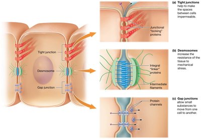
Cell junctions are connections between neighboring cells in a tissue, formed by integral proteins. They maintain tissue integrity and regulate movement between cells.
Tight (Occluding) Junctions: Locking proteins prevent passage of macromolecules between cells.
Desmosomes: Linker proteins distribute mechanical stress across cells.
Gap Junctions: Protein channels allow small substances to pass freely between cells.
Epithelial Tissues
Functions of Epithelial Tissue
Epithelial tissue is found on every external and internal body surface, acting as a barrier between the body and the environment. Its main functions include:
Protection: Shields underlying tissue from injury; produces keratin; rapid mitosis.
Immune Defenses: Contains immune cells.
Secretion: Forms glands that produce substances like sweat, oil, and hormones.
Transport: Selectively permeable barriers for substance movement.
Sensation: Supplied with nerves for detecting environmental changes.
Microscopic Appearance of Tissues
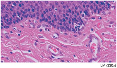
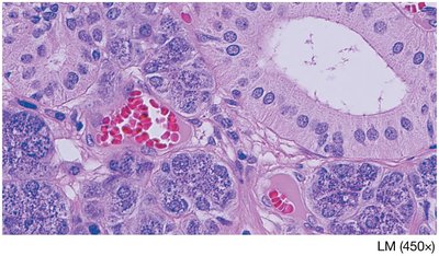
Histological sections are stained for microscopic study. Nuclei stain dark purple, ECM appears clear or lightly colored, and protein fibers appear wavy or as straight lines.
Components and Classification of Epithelia
The ECM of epithelia is located in the basement membrane, which anchors epithelial tissue to underlying connective tissue. The basement membrane consists of the basal lamina (collagen fibers and ground substance) and the reticular lamina (reticular fibers and ground substance).
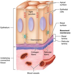
Epithelia are classified by:
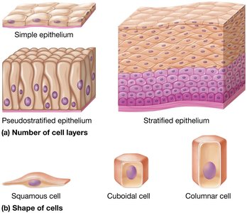
Number of Cell Layers: Simple (one layer), stratified (multiple layers), pseudostratified (appears multilayered but is single-layered).
Shape of Cells: Squamous (flattened), cuboidal (cube-shaped), columnar (tall and elongated).
Covering and Lining Epithelia
These epithelia form broad, flat sheets on inner and outer body surfaces. Simple epithelia are one cell layer thick and are specialized for diffusion, absorption, or secretion. Stratified epithelia are thicker and provide protection.
Simple Epithelia
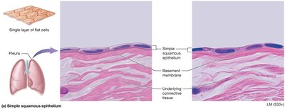
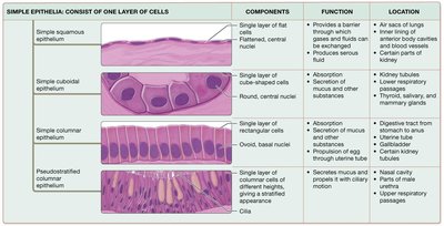
Simple Squamous: Single layer of flat cells; found in air sacs of lungs, serous membranes, and blood vessels.
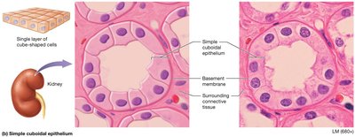
Simple Cuboidal: Single layer of cube-shaped cells; found in kidney tubules and glands.
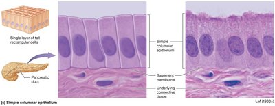
Simple Columnar: Single layer of tall cells; found in small intestine, uterine tube, kidney tubules, and glands.
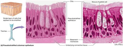
Pseudostratified Columnar: Appears stratified but is a single layer; found in respiratory passages and nasal cavities.
Transport Across Simple Epithelia
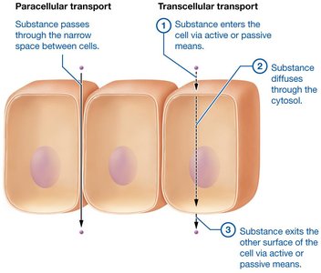
Paracellular Transport: Substances pass between cells.
Transcellular Transport: Substances move through cells (enter, diffuse, exit).
Stratified Epithelia
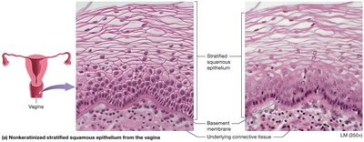
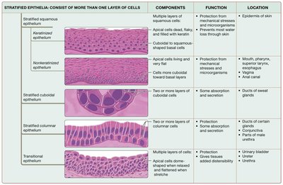
Stratified Squamous: Nonkeratinized type found in mouth, pharynx, esophagus, anus, and vagina.
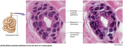
Stratified Cuboidal: Rare; found in ducts of sweat glands.
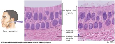
Stratified Columnar: Rare; found in ducts of salivary glands, male urethra, conjunctiva.
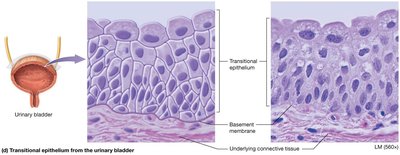
Transitional: Apical cells dome-shaped when relaxed, squamous when stretched; found in urinary tract.
Summary Tables of Epithelial Tissues
Type | Components | Function | Location |
|---|---|---|---|
Simple Squamous | Single layer of flat cells | Diffusion, filtration | Air sacs of lungs, lining of blood vessels |
Simple Cuboidal | Single layer of cube-shaped cells | Absorption, secretion | Kidney tubules, glands |
Simple Columnar | Single layer of tall cells | Absorption, secretion | Digestive tract, uterine tubes |
Pseudostratified Columnar | Single layer, appears stratified | Secretion, movement of mucus | Respiratory tract |
Type | Components | Function | Location |
|---|---|---|---|
Stratified Squamous | Multiple layers of squamous cells | Protection | Skin, mouth, esophagus, vagina |
Stratified Cuboidal | Two layers of cuboidal cells | Protection, secretion | Ducts of sweat glands |
Stratified Columnar | Columnar apical, cuboidal basal | Protection, secretion | Salivary gland ducts, male urethra |
Transitional | Multiple layers, dome-shaped apical cells | Distensibility | Urinary bladder, ureters |
Glandular Epithelia
Types of Glands
Exocrine Glands: Secrete products into ducts; local action (e.g., sweat, oil, salivary glands).
Endocrine Glands: Secrete hormones directly into blood; act on distant targets.
Exocrine Gland Structure
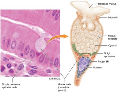
Unicellular: Goblet cells secrete mucus in digestive and respiratory tracts.
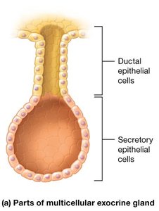
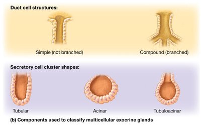
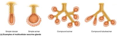
Multicellular: Classified by duct structure (simple or compound) and shape (tubular, acinar, tubuloacinar).
Additional info:
This summary covers the first half of Chapter 4 (Histology), focusing on tissue types, ECM, cell junctions, epithelial tissue structure and classification, and glandular epithelia. For a complete study guide, further sections on connective, muscle, and nervous tissues, as well as tissue repair and membranes, should be included.
