 Back
BackChapter 4: Skin and Body Membranes – Structure, Function, and Clinical Relevance
Study Guide - Smart Notes
 Tailored notes based on your materials, expanded with key definitions, examples, and context.
Skin and Body Membranes
Overview of Body Membranes
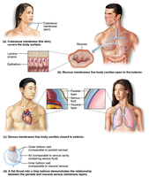
Body membranes are thin layers of tissue that cover surfaces, line body cavities, and form protective (often lubricating) sheets around organs. They are classified into epithelial membranes (cutaneous, mucous, and serous) and connective tissue membranes (synovial).
Cutaneous membrane: The skin; covers the body surface and is composed of keratinized stratified squamous epithelium and dense connective tissue.
Mucous membranes: Line body cavities open to the exterior (e.g., digestive, respiratory, urinary tracts); consist of epithelium on a loose connective tissue base (lamina propria).
Serous membranes: Line body cavities closed to the exterior (e.g., thoracic, abdominal cavities); composed of simple squamous epithelium on areolar connective tissue.
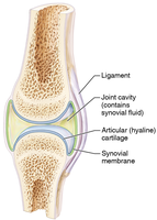
Synovial membranes: Composed entirely of connective tissue; line joint cavities and secrete synovial fluid for lubrication.
Functions of the Integumentary System
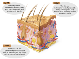
The integumentary system, primarily the skin, serves as the body's first line of defense and performs several vital functions.
Protection: Acts as a barrier against mechanical, chemical, microbial, and UV damage.
Regulation: Helps regulate body temperature and water loss.
Excretion: Excretes salts, water, and organic wastes via sweat.
Sensation: Contains sensory receptors for touch, pain, temperature, and pressure.
Synthesis: Synthesizes vitamin D when exposed to sunlight.
Detailed Functions and Mechanisms
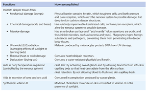
The skin accomplishes its protective and regulatory roles through specialized structures and biochemical processes.
Functions | How accomplished |
|---|---|
Protects deeper tissues from mechanical, chemical, and microbial damage | Keratinized cells, acid mantle, and immune cells provide barriers and defense |
Prevents UV damage | Melanin produced by melanocytes absorbs UV radiation |
Thermal regulation | Sweat glands and blood vessel dilation/constriction |
Excretion | Sweat glands remove urea, uric acid, and excess salts |
Vitamin D synthesis | UV light converts cholesterol derivatives to vitamin D |
Structure of the Skin
Layers of the Skin
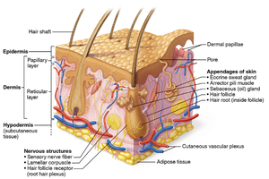
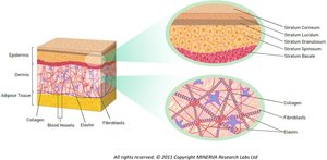
The skin is composed of three main layers: the epidermis, dermis, and hypodermis (subcutaneous tissue). Each layer has distinct structures and functions.
Epidermis: Outermost layer; avascular; composed of stratified squamous epithelium; contains keratinocytes, melanocytes, and other specialized cells.
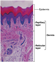
Dermis: Middle layer; vascular; contains connective tissue, blood vessels, nerves, hair follicles, and glands. Divided into papillary and reticular layers.
Hypodermis: Deepest layer; composed mainly of adipose tissue; anchors skin to underlying structures and provides insulation.
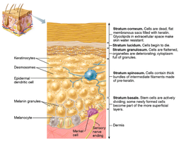
Epidermal Layers
The epidermis consists of several sublayers, each with specialized cells and functions:
Stratum corneum: Outermost, dead, keratin-filled cells; provides waterproof barrier.
Stratum lucidum: Present only in thick skin (palms, soles); clear, dead cells.
Stratum granulosum: Cells begin to die; contain granules of keratohyalin.
Stratum spinosum: Several layers of keratinocytes; provides strength and flexibility.
Stratum basale: Deepest layer; site of active cell division; contains melanocytes.
Color and Appendages of Skin
Skin Color
Skin color is determined by the amount and type of melanin, carotene, and hemoglobin present in the skin. Melanocytes in the stratum basale produce melanin, which protects against UV radiation.
Melanin: Brown-black pigment; more melanin = darker skin.
Carotene: Yellow-orange pigment from diet; accumulates in stratum corneum.
Hemoglobin: Red pigment in blood; gives pinkish hue to fair skin.
Skin Appendages
Skin appendages include cutaneous glands, hair and hair follicles, and nails. These structures arise from the epidermis and play roles in protection, sensation, and thermoregulation.
Cutaneous glands: Include sebaceous (oil) glands and sweat (sudoriferous) glands.
Hair and hair follicles: Provide protection, sensation, and help regulate body temperature.
Nails: Protect the distal phalanges and enhance fine touch.
Types of Sweat Glands
Eccrine glands: Widely distributed; secrete watery sweat for thermoregulation.
Apocrine glands: Found in axillary and genital areas; secrete thicker, milky sweat; become active at puberty.
Comparison: Eccrine glands are involved in temperature regulation and are active throughout life, while apocrine glands are associated with scent and become active during puberty.
Homeostatic Imbalances of the Skin
Infections, Allergies, and Cancer
The skin is susceptible to various disorders, including infections, allergies, and cancers.
Infections: Caused by bacteria, viruses, or fungi (e.g., impetigo, athlete's foot).
Allergies: Immune responses to substances like poison ivy or latex.
Skin cancer: Most common type of human cancer; includes basal cell carcinoma, squamous cell carcinoma, and melanoma.
Effects of Aging on Skin
With aging, the skin becomes thinner, drier, and less elastic. These changes increase sensitivity to cold, dryness, itchiness, and susceptibility to bruising. This is due to decreased collagen production, reduced oil gland activity, and thinning of the epidermis and dermis.
Burns and Hair Loss
Third-degree burns destroy both the epidermis and dermis, including hair follicles. Since hair follicles contain the stem cells necessary for hair regeneration, their destruction leads to permanent hair loss in the affected area.
Summary Table: Types of Body Membranes
Type | Location | Main Function |
|---|---|---|
Cutaneous | External body surface (skin) | Protection, sensation, thermoregulation |
Mucous | Lines cavities open to exterior | Secretion, absorption, protection |
Serous | Lines closed body cavities | Lubrication, reduces friction |
Synovial | Lines joint cavities | Secretes synovial fluid for lubrication |
