 Back
BackChapter 4: Tissue – The Living Fabric (ANP Study Notes)
Study Guide - Smart Notes
 Tailored notes based on your materials, expanded with key definitions, examples, and context.
Tissue: The Living Fabric
Introduction to Tissues
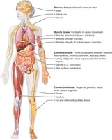
Tissues are groups of cells with similar structure and function, essential for maintaining homeostasis in the human body. The study of tissues is known as histology. There are four basic tissue types: epithelial, connective, muscle, and nervous tissue.
Epithelial Tissue: Covers surfaces and lines cavities; functions include protection, absorption, filtration, excretion, secretion, and sensory reception.
Connective Tissue: Supports, protects, binds other tissues, stores energy, and transports substances.
Muscle Tissue: Contracts to cause movement.
Nervous Tissue: Internal communication and control.
Histological Techniques
To study tissues under a microscope, samples must be fixed (preserved), sectioned (sliced thinly), and stained (colored for contrast). Light microscopy uses dyes, while electron microscopy uses heavy metal salts. Transmission electron microscopy (TEM) shows internal sections; scanning electron microscopy (SEM) shows surfaces.
Epithelial Tissue
General Characteristics
Epithelial tissue forms sheets of cells covering surfaces or lining cavities. It exists in two main forms: covering and lining epithelium (e.g., skin, lining of organs) and glandular epithelium (e.g., glands).
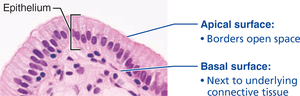
Polarity: Cells have distinct apical (top) and basal (bottom) surfaces.
Specialized Contacts: Cells are tightly joined by junctions (tight junctions, desmosomes).
Supported by Connective Tissue: Basement membrane reinforces the sheet.
Avascular but Innervated: No blood vessels, but supplied by nerves.
Regeneration: High capacity for renewal.
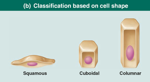
Classification of Epithelial Tissue
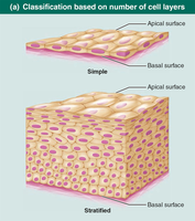
Epithelial tissues are classified by the number of cell layers and cell shape:
Simple Epithelium: Single layer; ideal for absorption, secretion, filtration.
Stratified Epithelium: Multiple layers; ideal for protection.
Squamous: Flattened, scale-like cells.
Cuboidal: Cube-shaped cells.
Columnar: Tall, column-like cells.
Types of Simple Epithelia
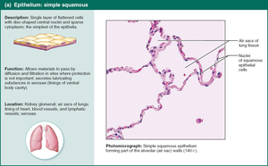
Simple Squamous Epithelium
Single layer of flat cells; allows rapid diffusion and filtration. Found in kidneys, lungs, lining of blood vessels (endothelium), and serous membranes (mesothelium).
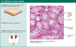
Simple Cuboidal Epithelium
Single layer of cube-shaped cells; functions in secretion and absorption. Found in kidney tubules and gland ducts.
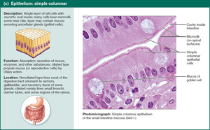
Simple Columnar Epithelium
Single layer of tall cells; may have microvilli or cilia. Functions in absorption and secretion of mucus and enzymes. Found in digestive tract, gallbladder, and uterine tubes.
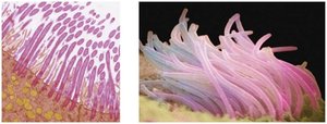
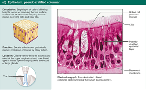
Pseudostratified Columnar Epithelium
Appears multi-layered but is actually a single layer; often ciliated. Functions in secretion and movement of mucus. Found in upper respiratory tract and large gland ducts.
Summary Table: Epithelial Tissue Types
Type | Structure | Main Function | Location |
|---|---|---|---|
Simple Squamous | Single layer, flat cells | Diffusion, filtration | Kidney, lungs, blood vessels |
Simple Cuboidal | Single layer, cube cells | Secretion, absorption | Kidney tubules, glands |
Simple Columnar | Single layer, tall cells | Absorption, secretion | Digestive tract, gallbladder |
Pseudostratified Columnar | Single layer, varied height | Mucus secretion, movement | Respiratory tract, glands |
Additional info:
Stratified epithelia (not shown in images) provide greater protection and are found in areas subject to abrasion, such as skin and mouth.
Transitional epithelium (not shown in images) lines urinary organs and can stretch to accommodate fluctuating volumes.
