 Back
BackChapter 4: Tissue—The Living Fabric (ANP Study Notes)
Study Guide - Smart Notes
 Tailored notes based on your materials, expanded with key definitions, examples, and context.
Tissue: The Living Fabric
Introduction to Tissues
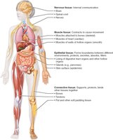
Tissues are groups of cells with similar structure and function, essential for maintaining homeostasis in the human body. The study of tissues is called histology. There are four basic tissue types: epithelial, connective, muscle, and nervous tissue.
Epithelial tissue: Covers surfaces and lines cavities; functions in protection, absorption, filtration, excretion, secretion, and sensory reception.
Connective tissue: Supports, protects, binds other tissues, stores energy, and transports substances.
Muscle tissue: Contracts to produce movement.
Nervous tissue: Initiates and transmits electrical impulses for communication.
4.1 Tissue Preparation for Microscopy
Microscopy Techniques
To study tissues under a microscope, samples must be fixed (preserved), sectioned (sliced thinly), and stained (colored for contrast). Light microscopy uses dyes, while electron microscopy uses heavy metal salts. Transmission electron microscopy (TEM) shows internal sections, and scanning electron microscopy (SEM) shows surfaces.
4.2 Epithelial Tissue
Definition and Functions
Epithelial tissue is a sheet of cells covering body surfaces or lining cavities. It exists in two main forms: covering and lining epithelium (e.g., skin, lining of organs) and glandular epithelium (forms glands). Main functions include protection, absorption, filtration, excretion, secretion, and sensory reception.
Special Characteristics of Epithelial Tissue
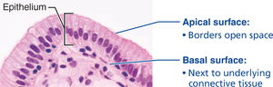
Polarity: Cells have distinct apical (top) and basal (bottom) surfaces. The apical surface may have microvilli; the basal surface attaches to the basal lamina.
Specialized contacts: Cells are tightly joined by junctions (tight junctions, desmosomes).
Supported by connective tissue: All epithelial sheets rest on a basement membrane (basal and reticular lamina).
Avascular but innervated: No blood vessels, but supplied by nerves; nutrients diffuse from underlying connective tissue.
Regeneration: High capacity for renewal, especially in areas exposed to friction or hostile environments.
Classification of Epithelial Tissue
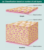
Epithelial tissues are classified by number of cell layers and cell shape:
Simple epithelium: Single layer; ideal for absorption, secretion, filtration.
Stratified epithelium: Multiple layers; ideal for protection in high-abrasion areas.
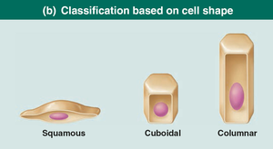
Cell shapes: Squamous (flat), cuboidal (cube-shaped), columnar (tall).
Types of Simple Epithelia
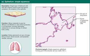
Simple Squamous Epithelium
Single layer of flat cells; allows rapid diffusion and filtration. Found in kidneys, lungs, lining of blood vessels (endothelium), and serous membranes (mesothelium).
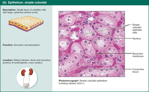
Simple Cuboidal Epithelium
Single layer of cube-shaped cells; functions in secretion and absorption. Found in kidney tubules, ducts, and glands.
Simple Columnar Epithelium
Single layer of tall cells; may have microvilli or cilia. Functions in absorption and secretion of mucus, enzymes, and other substances. Found in digestive tract, gallbladder, bronchi, and uterine tubes.

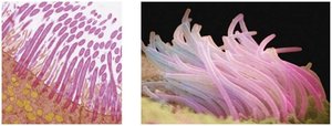
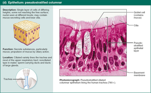
Pseudostratified Columnar Epithelium
Appears multi-layered but is actually a single layer; often ciliated. Functions in secretion and movement of mucus. Found in upper respiratory tract, ducts of large glands, and tubules in testes.
Types of Stratified Epithelia
Stratified squamous epithelium: Most common; protects against abrasion. Keratinized (skin) and nonkeratinized (moist linings).
Stratified cuboidal and columnar epithelium: Rare; found in sweat glands, mammary glands, pharynx, male urethra.
Transitional epithelium: Lines urinary organs; cells change shape to allow stretching.
Glandular Epithelia
A gland is one or more cells that secrete an aqueous fluid. Glands are classified by site of product release (endocrine or exocrine) and number of cells (unicellular or multicellular).
Endocrine glands: Ductless; secrete hormones into interstitial fluid and blood.
Exocrine glands: Secrete products into ducts or onto surfaces; include sweat, oil, and salivary glands.
Unicellular exocrine glands: Goblet cells and mucous cells; produce mucin.
Multicellular exocrine glands: Composed of duct and secretory unit; classified by structure and mode of secretion (merocrine, holocrine, apocrine).
4.3 Connective Tissue
Definition and Functions
Connective tissue is the most abundant and widely distributed tissue type. Functions include binding, support, protection, insulation, energy storage, and transport. Four main classes: connective tissue proper, cartilage, bone, and blood.
Common Characteristics
Extracellular matrix: Nonliving material surrounding cells; allows tissue to withstand tension and abuse.
Common origin: All connective tissues arise from embryonic mesenchyme.
Structural Elements
Ground substance: Gel-like material; contains interstitial fluid, cell adhesion proteins, and proteoglycans (e.g., chondroitin sulfate, hyaluronic acid).
Fibers: Collagen (strong), elastic (stretchy), reticular (supportive networks).
Cells: Immature "-blast" cells (secrete matrix), mature "-cyte" cells (maintain matrix), adipocytes (fat storage), leukocytes (immune response), mast cells (inflammation), macrophages (phagocytosis).
Types of Connective Tissue Proper
Loose connective tissue: Areolar (packing material), adipose (fat storage), reticular (support for blood cells).
Dense connective tissue: Dense regular (tendons, ligaments), dense irregular (dermis, organ capsules), elastic (arteries, vertebral ligaments).
Cartilage
Cartilage is tough yet flexible, avascular, and lacks nerve fibers. Types include hyaline (most abundant), elastic (ears, epiglottis), and fibrocartilage (intervertebral discs, knee).
Bone (Osseous Tissue)
Bone supports and protects, stores fat, and synthesizes blood cells. Contains more collagen and inorganic calcium salts. Osteoblasts produce matrix; osteocytes maintain it.
Blood
Blood is a fluid connective tissue; consists of cells in plasma. Functions in transport of nutrients, wastes, gases, and other substances.
4.4 Muscle Tissue
Types of Muscle Tissue
Skeletal muscle: Voluntary, striated, multinucleated; moves bones.
Cardiac muscle: Involuntary, striated, one nucleus per cell, intercalated discs; found in heart.
Smooth muscle: Involuntary, non-striated, spindle-shaped; found in walls of hollow organs.
4.5 Nervous Tissue
Structure and Function
Nervous tissue is the main component of the nervous system. It consists of neurons (transmit electrical signals) and supporting glial cells (support, insulate, protect neurons).
4.6 Membranes
Types of Membranes
Cutaneous membrane: Skin; dry, keratinized stratified squamous epithelium attached to connective tissue.
Mucous membrane: Lines cavities open to exterior; moist, may secrete mucus.
Serous membrane: Lines closed ventral cavities; moist, simple squamous epithelium (mesothelium) on areolar connective tissue.
4.7 Tissue Repair
Steps in Tissue Repair
Inflammation: Release of chemicals, dilation of blood vessels, clotting.
Organization: Blood clot replaced by granulation tissue, regeneration begins, fibroblasts bridge gap.
Regeneration and fibrosis: Scab detaches, epithelium regenerates, scar tissue forms.
Regenerative Capacity
High: Epithelial, bone, areolar, dense irregular, blood-forming tissue.
Moderate: Smooth muscle, dense regular connective tissue.
Low/None: Cardiac muscle, nervous tissue.
Developmental Aspects of Tissues
Primary Germ Layers
Ectoderm: Forms nerve tissue.
Mesoderm: Forms muscle and connective tissue.
Endoderm: Contributes to epithelial tissue.
Tissues function well in youth; aging leads to thinning epithelia, less efficient repair, atrophy, and increased cancer risk.
