 Back
BackChapter 4: Tissue—The Living Fabric (Epithelial and Connective Tissues)
Study Guide - Smart Notes
 Tailored notes based on your materials, expanded with key definitions, examples, and context.
Tissue: The Living Fabric
Introduction to Tissues
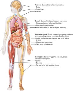
Tissues are groups of cells similar in structure that perform common or related functions. The study of tissues is called histology. There are four primary tissue types in the human body: epithelial, connective, muscle, and nervous tissue.
Epithelial tissue: Covers surfaces and lines cavities.
Connective tissue: Supports, protects, and binds other tissues.
Muscle tissue: Contracts to produce movement.
Nervous tissue: Enables internal communication.
Epithelial Tissue
General Characteristics
Epithelial tissue is a sheet of cells that covers body surfaces or lines body cavities. It has two main forms:
Covering and lining epithelium: Forms the outer layer of the skin and lines open cavities and organs.
Glandular epithelium: Forms glands that secrete substances (e.g., salivary glands).
Main functions include protection, absorption, filtration, excretion, secretion, and sensory reception.
Special Characteristics of Epithelial Tissue
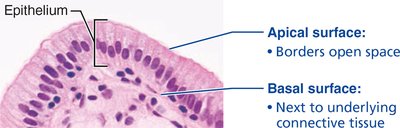
Polarity: Cells have an apical (top) surface and a basal (bottom) surface.
Specialized contacts: Cells are tightly joined by junctions (tight junctions and desmosomes).
Supported by connective tissue: The basement membrane anchors epithelium to underlying connective tissue.
Avascular but innervated: Contains no blood vessels but is supplied by nerve fibers; nutrients diffuse from underlying tissues.
Regeneration: High capacity for renewal, especially in areas exposed to friction.
Classification of Epithelial Tissue
Epithelial tissues are classified by the number of cell layers and the shape of the cells in the apical layer.
Number of layers:
Simple epithelium: Single cell layer (for absorption, secretion, filtration).
Stratified epithelium: Two or more layers (for protection).
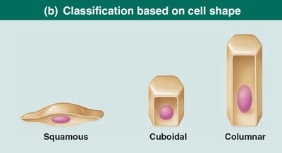
Cell shape:
Squamous: Flat and scale-like.
Cuboidal: Cube-shaped.
Columnar: Tall and column-like.

Types of Epithelial Tissue
Simple Squamous Epithelium
Description: Single layer of flattened cells with disc-shaped central nuclei.
Function: Allows materials to pass by diffusion and filtration; secretes lubricating substances.
Location: Kidney glomeruli, air sacs of lungs, lining of heart, blood vessels, lymphatic vessels, serosae.

Simple Cuboidal Epithelium
Description: Single layer of cube-like cells with large, spherical nuclei.
Function: Secretion and absorption.
Location: Kidney tubules, ducts and secretory portions of small glands, ovary surface.

Simple Columnar Epithelium
Description: Single layer of tall cells; may have microvilli or cilia; may contain mucus-secreting goblet cells.
Function: Absorption; secretion of mucus, enzymes, and other substances; ciliated type propels mucus.
Location: Nonciliated: digestive tract; ciliated: small bronchi, uterine tubes, some regions of uterus.

Pseudostratified Columnar Epithelium
Description: Single layer of cells of differing heights; may be ciliated; appears stratified but is not.
Function: Secretion, particularly of mucus; propulsion of mucus by ciliary action.
Location: Ciliated: trachea, upper respiratory tract; nonciliated: sperm-carrying ducts, ducts of large glands.

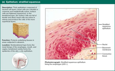
Stratified Squamous Epithelium
Description: Thick membrane composed of several cell layers; basal cells are cuboidal or columnar, surface cells are flattened.
Function: Protects underlying tissues in areas subject to abrasion.
Location: Nonkeratinized: moist linings of esophagus, mouth, vagina; keratinized: epidermis of skin.
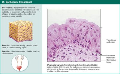
Transitional Epithelium
Description: Resembles both stratified squamous and stratified cuboidal; basal cells are cuboidal or columnar; surface cells dome-shaped or squamous-like depending on degree of organ stretch.
Function: Stretches readily, permits stored urine to distend urinary organ.
Location: Lines the ureters, bladder, and part of the urethra.
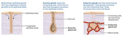
Glandular Epithelia
Gland: One or more cells that make and secrete an aqueous fluid called a secretion.
Classification by site of product release:
Endocrine: Ductless glands; secrete hormones into blood or lymph.
Exocrine: Secrete products onto body surfaces or into body cavities via ducts (e.g., sweat, oil, salivary glands).
Classification by number of cells:
Unicellular: Goblet cells (produce mucin).
Multicellular: Composed of a duct and secretory unit; classified by structure and mode of secretion.
Modes of Secretion in Multicellular Exocrine Glands
Merocrine: Secrete products by exocytosis (e.g., sweat glands, pancreas).
Holocrine: Accumulate products until cell ruptures (e.g., sebaceous glands).
Apocrine: Accumulate products, but only apex ruptures (e.g., mammary glands).
Connective Tissue
General Features
Connective tissue is the most abundant and widely distributed tissue type. Its major functions include binding and support, protection, insulation, storing reserve fuel, and transporting substances (e.g., blood).
Four main classes:
Connective tissue proper
Cartilage
Bone
Blood
Common Characteristics
Extracellular matrix: Nonliving material that separates living cells; supports cells so they can bear weight and withstand tension.
Common origin: All connective tissues arise from mesenchyme (embryonic tissue).
Structural Elements
Ground substance: Unstructured material that fills space between cells; medium for diffusion of nutrients and waste.
Fibers: Provide support; three types:
Collagen fibers: Strongest, most abundant; high tensile strength.
Elastic fibers: Long, thin, allow for stretch and recoil.
Reticular fibers: Short, fine, highly branched; form networks for support.
Cells: Each major class has a resident cell type in immature (-blast) and mature (-cyte) forms (e.g., fibroblasts/fibrocytes, chondroblasts/chondrocytes, osteoblasts/osteocytes).
Types of Connective Tissue
Tissue Class | Subclasses | Cells | Matrix | General Features |
|---|---|---|---|---|
Connective Tissue Proper | Loose (areolar, adipose, reticular); Dense (regular, irregular, elastic) | Fibroblasts, fibrocytes, adipocytes, defense cells | Gel-like ground substance; all three fiber types | Binding tissue, resists tension, energy storage |
Cartilage | Hyaline, elastic, fibrocartilage | Chondroblasts, chondrocytes | Gel-like ground substance; collagen, elastic fibers | Resists compression, supports, avascular |
Bone | Compact, spongy | Osteoblasts, osteocytes | Calcified matrix; collagen fibers | Support, protection, stores fat, blood cell formation |
Blood | --- | Red and white blood cells, platelets | Plasma (fluid); no fibers | Transport of gases, nutrients, wastes |
Other Cell Types in Connective Tissue
Adipocytes: Store energy as fat.
Leukocytes (white blood cells): Respond to injury and infection.
Mast cells: Mediate inflammation (release histamine, heparin).
Macrophages: Phagocytize foreign materials and dead cells.
Muscle Tissue
General Features
Muscle tissue is highly vascularized and responsible for movement. Muscle cells contain myofilaments (actin and myosin) for contraction. There are three types:
Skeletal muscle: Voluntary movement, attached to bones.
Cardiac muscle: Involuntary, found in the heart, allows coordinated contraction via intercalated discs.
Smooth muscle: Involuntary, found in walls of hollow organs (e.g., intestines, blood vessels).
Nervous Tissue
General Features
Nervous tissue is the main component of the nervous system (brain, spinal cord, nerves). It regulates and controls body functions. Two main cell types:
Neurons: Respond to stimuli and transmit electrical signals via dendrites and axons.
Supporting cells (neuroglia): Support, insulate, and protect neurons.
