 Back
BackChapter 4: Tissue—The Living Fabric (Marieb Human Anatomy & Physiology, 12th Edition)
Study Guide - Smart Notes
 Tailored notes based on your materials, expanded with key definitions, examples, and context.
Chapter 4: Tissue—The Living Fabric
Introduction to Tissues
Tissues are groups of cells similar in structure that perform a common or related function. The study of tissues is called histology. Understanding tissues is fundamental to comprehending how organs and organ systems function in the human body.
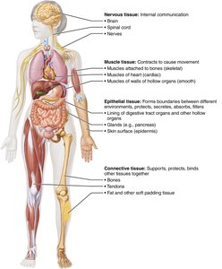
Four basic tissue types: Epithelial, Connective, Muscle, and Nervous tissues
Each tissue type has distinct roles in the body, contributing to structure, protection, movement, and communication.

Overview of the Four Basic Tissue Types
Epithelial Tissue: Forms boundaries between different environments, protects, secretes, absorbs, and filters. Found in the skin surface (epidermis) and lining of digestive tract organs.
Connective Tissue: Supports, protects, and binds other tissues together. Examples include bones, tendons, and fat.
Muscle Tissue: Contracts to cause movement. Includes skeletal muscles, cardiac muscle, and smooth muscle in organ walls.
Nervous Tissue: Enables internal communication through the brain, spinal cord, and nerves.
Epithelial Tissue
General Characteristics of Epithelial Tissue
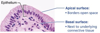
Epithelial tissue covers body surfaces, lines cavities, and forms glands. It is characterized by closely packed cells with minimal extracellular material, polarity (apical and basal surfaces), and a high capacity for regeneration.
Polarity: Has an apical surface (exposed to exterior or cavity) and a basal surface (attached to underlying connective tissue).
Specialized contacts: Cells are joined by tight junctions and desmosomes.
Supported by connective tissue: The basal surface rests on a basement membrane.
Avascular but innervated: Contains no blood vessels but is supplied by nerve fibers.
High regenerative capacity: Rapidly replaces lost cells by cell division.
Classification of Epithelia
Epithelia are classified based on the number of cell layers and the shape of the cells.
Number of layers:
Simple epithelium: Single cell layer (for absorption, secretion, filtration).
Stratified epithelium: Two or more layers (for protection).
Cell shape:
Squamous: Flattened and scale-like.
Cuboidal: Boxlike, as tall as they are wide.
Columnar: Tall and column shaped.

Types of Epithelial Tissue
Simple Squamous Epithelium:
Description: Single layer of flattened cells with disc-shaped central nuclei.
Function: Allows diffusion and filtration; secretes lubricating substances.
Location: Air sacs of lungs, lining of heart, blood vessels.

Simple Cuboidal Epithelium:
Description: Single layer of cube-like cells with large, spherical nuclei.
Function: Secretion and absorption.
Location: Kidney tubules, ducts, and secretory portions of small glands.

Simple Columnar Epithelium:
Description: Single layer of tall cells; may have microvilli or cilia.
Function: Absorption; secretion of mucus, enzymes, and other substances.
Location: Nonciliated type lines digestive tract; ciliated type lines small bronchi, uterine tubes.

Pseudostratified Columnar Epithelium:
Description: Single layer of cells of differing heights; may contain mucus-secreting cells and bear cilia.
Function: Secretes substances, particularly mucus; propulsion of mucus by ciliary action.
Location: Ciliated type in trachea and upper respiratory tract.

Stratified Squamous Epithelium:
Description: Thick membrane composed of several cell layers; basal cells are cuboidal or columnar, surface cells are flattened.
Function: Protects underlying tissues in areas subject to abrasion.
Location: Nonkeratinized type forms moist linings of esophagus, mouth, vagina; keratinized type forms epidermis of skin.

Transitional Epithelium:
Description: Resembles both stratified squamous and cuboidal; surface cells dome-shaped or squamous depending on degree of organ stretch.
Function: Stretches readily, permits stored urine to distend urinary organ.
Location: Lines ureters, bladder, and part of urethra.

Glandular Epithelia
Glandular epithelia form glands that secrete substances onto surfaces or into the bloodstream. Glands are classified as endocrine (ductless, secrete hormones) or exocrine (secrete via ducts).
Formation: Multicellular glands form by invagination (inward growth) of epithelial sheets.
Exocrine glands: Retain connecting ducts; secrete onto epithelial surfaces (e.g., sweat, salivary glands).
Endocrine glands: Lose ducts during development; secrete hormones into blood (e.g., thyroid, pituitary).

Unicellular Exocrine Glands: Goblet Cells
Goblet cells are the only important unicellular exocrine glands in humans. They produce mucin, which dissolves in water to form mucus, a protective, lubricating secretion.
Location: Found in epithelial linings of intestinal and respiratory tracts.
Structure: Characterized by an abundance of secretory vesicles containing mucin.

Types of Multicellular Exocrine Glands
Multicellular exocrine glands are classified by duct structure (simple or compound) and secretory units (tubular, alveolar, or tubuloalveolar).
Structure | Simple Duct | Compound Duct |
|---|---|---|
Tubular | Intestinal glands | Duodenal glands of small intestine |
Alveolar | No important example in humans | Mammary glands |
Tubuloalveolar | Stomach (gastric) glands | Salivary glands |

Modes of Secretion in Exocrine Glands
Merocrine: Secrete products by exocytosis (e.g., sweat, pancreas).
Holocrine: Entire secretory cell ruptures, releasing secretions and cell fragments (e.g., sebaceous glands).

Connective Tissue
General Features of Connective Tissue
Connective tissue is the most abundant and widely distributed tissue in the body. It supports, protects, and binds other tissues. All connective tissues share three basic components: cells, fibers, and ground substance (together forming the extracellular matrix).
Major classes: Connective tissue proper, cartilage, bone, and blood.
Functions: Binding and support, protection, insulation, storing reserve fuel, and transporting substances.

Components of Connective Tissue
Fibers: Collagen (strength), elastic (stretch), and reticular (support).
Ground substance: Unstructured material that fills the space between cells and fibers.
Cells: Fibroblasts (in connective tissue proper), chondroblasts (in cartilage), osteoblasts (in bone), and hematopoietic stem cells (in blood).

Classification of Connective Tissue
Connective tissue is classified into connective tissue proper (loose and dense), cartilage, bone, and blood. Each class has unique cells, fibers, and ground substance composition.
Loose connective tissue: Areolar, adipose, reticular
Dense connective tissue: Regular, irregular, elastic
Cartilage: Hyaline, elastic, fibrocartilage
Bone
Blood

Areolar Connective Tissue
Description: Gel-like matrix with all three fiber types; cells include fibroblasts, macrophages, mast cells, and some white blood cells.
Function: Wraps and cushions organs; plays a role in inflammation; holds and conveys tissue fluid.
Location: Widely distributed under epithelia of body, forms lamina propria of mucous membranes.

Adipose Connective Tissue
Description: Matrix as in areolar, but very sparse; closely packed adipocytes.
Function: Provides reserve food fuel; insulates against heat loss; supports and protects organs.
Location: Under skin in subcutaneous tissue; around kidneys and eyeballs; within abdomen; in breasts.

Summary Table: Classes of Connective Tissues
Tissue Class and Example | Subclasses | Cells | Matrix | General Features |
|---|---|---|---|---|
Connective Tissue Proper | Loose (areolar, adipose, reticular); Dense (regular, irregular, elastic) | Fibroblasts, fibrocytes, defense cells, adipocytes | Gel-like ground substance; all three fiber types | Six different types; vary in density and types of fibers |
Cartilage | Hyaline, elastic, fibrocartilage | Chondroblasts, chondrocytes | Gel-like ground substance; fibers: collagen, elastic fibers in some | Resists compression; functions to cushion and support body structures |
Bone Tissue | Compact bone | Osteoblasts, osteocytes | Gel-like ground substance calcified with inorganic salts; fibers: collagen | Hard tissue that resists both compression and tension; functions in support |
Blood | --- | Red blood cells, white blood cells, platelets | Plasma (no fibers) | A fluid tissue; functions to carry O2, CO2, nutrients, wastes, and other substances |
Additional info:
This summary covers the main types and characteristics of epithelial and connective tissues, as well as their classification and functions. For a complete understanding, students should also study muscle and nervous tissues, tissue repair, and developmental aspects of tissues as outlined in the full chapter.
