 Back
BackChapter 5: The Integumentary System – Anatomy & Physiology Study Guide
Study Guide - Smart Notes
 Tailored notes based on your materials, expanded with key definitions, examples, and context.
Chemical barrier: Antimicrobial proteins, acid mantle, melanin.
Physical barrier: Keratinized cells, glycolipids.
Biological barrier: Dendritic cells, macrophages, DNA absorbs UV.
Body Temperature RegulationThe Integumentary System
Overview
The integumentary system is the body's largest organ system, consisting of the skin and its derivatives: hair, nails, sweat glands, and sebaceous glands. It serves as a protective barrier, regulates temperature, and provides sensory information.
Components: Skin, hair, nails, sweat glands, sebaceous (oil) glands
Main Functions: Protection, regulation, sensation, metabolic activity, blood reservoir, excretion
Structure of Skin
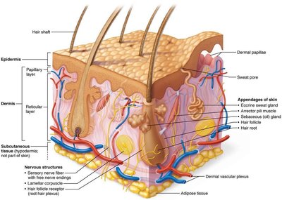
Skin Layers
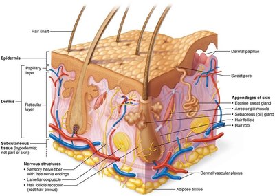
The skin is composed of three main layers, each with distinct functions and structures.
Epidermis: The superficial, avascular layer made of epithelial tissue.
Dermis: The deeper, vascular layer composed of connective tissue.
Hypodermis (Subcutaneous layer): Not technically part of the skin, but anchors it to underlying structures; mainly adipose tissue for shock absorption and insulation.
Epidermis
Cell Types in the Epidermis
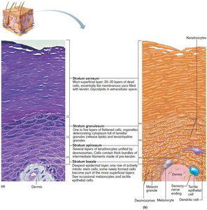
The epidermis is primarily made of keratinized stratified squamous epithelium and contains four main cell types:
Keratinocytes: Produce keratin, the protein that gives skin its strength and waterproofing.
Melanocytes: Produce melanin pigment, which protects against UV radiation.
Dendritic (Langerhans) cells: Immune cells that patrol the epidermis.
Tactile (Merkel) cells: Sensory receptors for touch.
Layers of the Epidermis
The epidermis consists of four or five distinct layers (strata), depending on skin thickness:
Stratum basale: Deepest layer; single row of stem cells; active mitosis; contains melanocytes.
Stratum spinosum: Several layers thick; contains keratinocytes, melanosomes, dendritic cells.
Stratum granulosum: Four to six layers; keratinization begins; cells flatten and die.
Stratum lucidum: Only in thick skin (palms, soles); two to three rows of clear, dead keratinocytes.
Stratum corneum: 20–30 rows of dead, keratinized cells; provides protection and prevents water loss.
Dermis
Structure and Layers
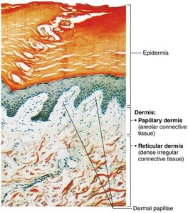
The dermis is a strong, flexible connective tissue layer containing nerves, blood vessels, lymphatics, hair follicles, and glands. It is divided into two layers:
Papillary layer: Superficial; areolar connective tissue; contains dermal papillae with capillaries and touch receptors.
Reticular layer: Deep; dense irregular connective tissue; provides strength and elasticity; contains cleavage and flexure lines.
Dermal Modifications
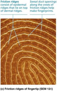
Friction ridges: Enhance grip and create fingerprints.
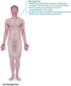
Cleavage (tension) lines: Parallel collagen fibers; important for surgical incisions.
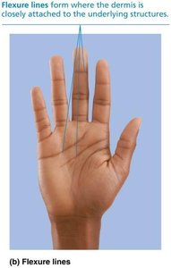
Flexure lines: Dermal folds near joints; visible on hands and feet.
Clinical Note: Stretch Marks and Blisters
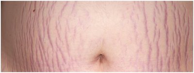
Striae: Silvery white scars from dermal tears (stretch marks).
Blisters: Fluid-filled pockets separating epidermal and dermal layers.
Skin Color
Pigments
Three pigments contribute to skin color:
Melanin: Produced by melanocytes; shields DNA from UV; color varies from yellow to black.
Carotene: Yellow to orange pigment; accumulates in stratum corneum and hypodermis.
Hemoglobin: Red pigment in blood; gives pinkish hue to fair skin.
Clinical Note: Skin Color Changes
Cyanosis: Blue skin; low oxygenation.
Pallor: Pale skin; anemia or low blood pressure.
Erythema: Redness; fever, inflammation.
Jaundice: Yellow skin; liver disorder.
Bruises: Clotted blood beneath skin.
Hair
Structure and Function
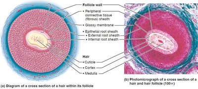
Hair consists of dead, keratinized cells and is produced by hair follicles. Functions include protection, sensation, and regulation.
Regions: Shaft (above skin), root (within skin).
Parts of shaft: Medulla (core), cortex (middle), cuticle (outer layer).
Pigmentation: Melanocytes produce melanin; color varies by type and amount.
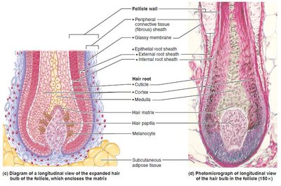
Hair Follicle Structure
Hair bulb: Expanded deep end; contains matrix for cell division.
Arrector pili: Smooth muscle causing "goose bumps".
Hair papilla: Dermal tissue supplying nutrients.
Types and Growth
Vellus hair: Fine, pale body hair.
Terminal hair: Coarse, long hair (scalp, eyebrows, pubic regions).
Growth: Influenced by nutrition and hormones; follicles cycle between active and regressive phases.
Clinical Note: Hair Disorders
Hirsutism: Excessive hairiness due to androgen excess.
Alopecia: Hair thinning after age 40.
Male pattern baldness: Genetic, influenced by DHT.
Telogen effluvium: Abrupt hair thinning due to stress or illness.
Nails
Structure and Function
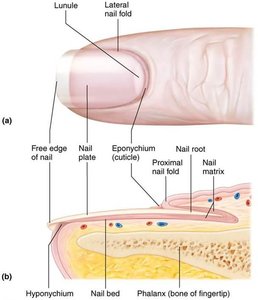
Nails are scale-like modifications of the epidermis containing hard keratin. They protect the distal phalanges and aid in manipulation.
Parts: Free edge, nail plate, root, nail bed, nail matrix (growth area).
Nail folds: Skin folds overlapping nail border.
Eponychium: Cuticle.
Hyponychium: Area under free edge.
Lunule: White, thickened area of matrix.
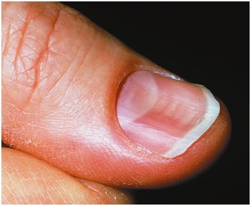
Clinical Note: Nail Disorders
Yellow nails: May indicate respiratory or thyroid disorder.
Thickened yellow nails: Fungal infection.
Koilonychia: Spoon-shaped nails; iron deficiency.
Beau’s lines: Horizontal lines; severe illness.
Cutaneous Glands
Sweat Glands
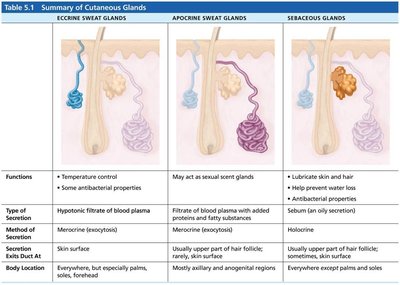
Eccrine (merocrine) glands: Most numerous; thermoregulation; secrete watery sweat.
Apocrine glands: Found in axillary/anogenital areas; secrete viscous sweat; function as scent glands.
Modified apocrine glands: Ceruminous (earwax) and mammary (milk) glands.
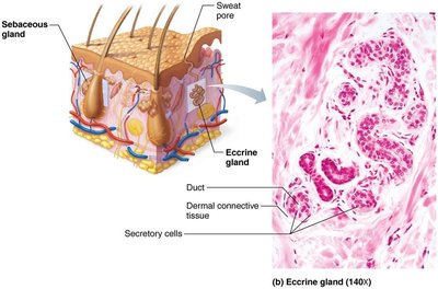
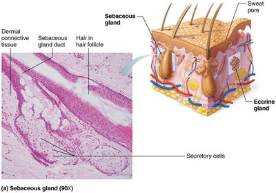
Sebaceous (Oil) Glands
Location: Everywhere except palms and soles; usually associated with hair follicles.
Function: Secrete sebum (oily, bactericidal); softens skin and hair.
Activation: Stimulated by hormones, especially androgens.
Summary Table: Cutaneous Glands
Gland Type | Function | Secretion | Location |
|---|---|---|---|
Eccrine Sweat | Temperature control, antibacterial | Watery, hypotonic filtrate | Palms, soles, forehead |
Apocrine Sweat | Sexual scent | Viscous, fatty/protein-rich | Axillary, anogenital |
Sebaceous | Lubricate, antibacterial | Oily sebum | Everywhere except palms/soles |
Clinical Note: Gland Disorders
Acne: Infectious inflammation of sebaceous glands.
Whiteheads/Blackheads: Blocked sebaceous glands.
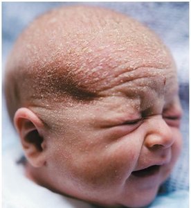
Cradle cap (seborrhea): Overactive sebaceous glands in infants.
Functions of Skin
Protection
The skin acts as a barrier against physical, chemical, and biological threats.
Insensible perspiration: Unnoticeable sweat (500 ml/day).
Sensible perspiration: Noticeable sweat (up to 12 L/day) for cooling.
Cold response: Dermal vessels constrict to conserve heat.
Cutaneous Sensations
Exteroreceptors: Respond to external stimuli (touch, temperature).
Free nerve endings: Sense pain.
Metabolic Functions
Vitamin D synthesis: Needed for calcium absorption.
Keratinocytes: Disarm carcinogens, activate hormones, produce collagenase.
Blood Reservoir
Skin holds up to 5% of blood volume.
Vessels can constrict to redirect blood.
Excretion
Excretes nitrogenous wastes: Ammonia, urea, uric acid.
Sweating: Causes salt and water loss.
Skin Cancer and Burns
Skin Cancer
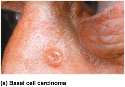
Basal cell carcinoma: Most common, least malignant; arises from stratum basale.
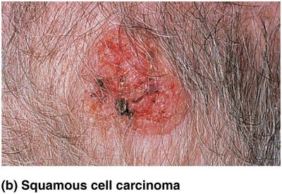
Squamous cell carcinoma: Second most common; arises from stratum spinosum; can metastasize.
Melanoma: Most dangerous; cancer of melanocytes; highly metastatic.
Burns
First-degree: Epidermal damage only; redness, pain.
Second-degree: Epidermal and upper dermal damage; blisters.
Third-degree: Entire skin thickness; gray-white, red, or black; no pain (nerve destruction); requires grafting.
Rule of Nines: Used to estimate fluid loss and burn severity.
Developmental Aspects of the Integumentary System
Fetal Development
Lanugo coat: Delicate hairs in 5th/6th month.
Vernix caseosa: Sebaceous secretion protecting fetal skin.
Infancy to Adulthood
Skin thickens, fat accumulates, gland activity increases.
Optimal appearance in 20s/30s; aging leads to thinning, dryness, wrinkles, increased cancer risk.
Aging Skin
Decreased replacement, elasticity, fat, and immune cells.
Hair thinning.
Prevention: UV protection, nutrition, hydration, hygiene.
