 Back
BackChapter 5: The Integumentary System – Structure, Function, and Clinical Relevance
Study Guide - Smart Notes
 Tailored notes based on your materials, expanded with key definitions, examples, and context.
The Skin and the Hypodermis
Overview and Structure
The skin is the largest organ of the human body, accounting for approximately 7% of body weight. It varies in thickness from 1.5 to 4.4 mm and is divided into two primary layers: the epidermis and the dermis. Beneath these lies the hypodermis, which, although not technically part of the integumentary system, shares many properties with the skin.
Epidermis: The superficial layer, composed of keratinized stratified squamous epithelium.
Dermis: The deeper layer, made of strong, flexible connective tissue.
Hypodermis: Composed of areolar and adipose tissues; functions in insulation and anchoring skin to underlying structures.
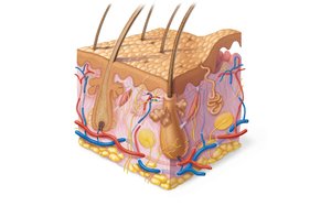
Functions of the Skin and Hypodermis
The skin and hypodermis perform several vital functions:
Protection: Cushions organs and protects against mechanical injury, chemicals, water loss, and UV radiation.
Body Temperature Regulation: Capillary networks and sweat glands regulate heat loss.
Excretion: Sweat removes urea, salts, and water.
Vitamin D Production: Epidermal cells synthesize vitamin D using UV radiation.
Sensory Reception: Contains sense organs associated with nerve endings.
Epidermis
Cell Types in the Epidermis
The epidermis is composed of four main cell types:
Keratinocytes: Most abundant; produce keratin, antibiotics, and enzymes. Located primarily in the stratum spinosum.
Melanocytes: Located in the basal layer; manufacture and secrete the pigment melanin.
Tactile Epithelial Cells: Located in the basal layer; attached to sensory nerve endings.
Dendritic Cells: Located in the stratum spinosum; function as macrophage-like cells in the immune system.
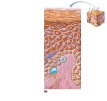
Layers of the Epidermis
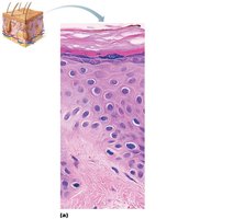
The epidermis consists of several distinct layers, each with unique characteristics:
Stratum Basale: Deepest layer; actively dividing cells; contains melanocytes and tactile epithelial cells.
Stratum Spinosum: Several layers of keratinocytes unified by desmosomes; contains dendritic cells.
Stratum Granulosum: One to five layers of flattened cells; contains keratohyaline and lamellar granules.
Stratum Lucidum: Present only in thick skin (palms, soles); a few rows of flat, dead keratinocytes.
Stratum Corneum: Most superficial; 20–30 layers of dead cells filled with keratin; provides protection against abrasion and penetration.
Dermis
Structure and Layers
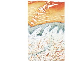
The dermis is the second major region of the skin, composed of strong, flexible connective tissue. It is richly supplied with blood vessels and nerves and consists of two layers:
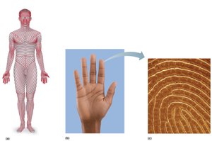
Papillary Dermis: Includes dermal papillae, which increase surface area for exchange of gases, nutrients, and wastes. Dermal papillae lie on dermal ridges, forming epidermal ridges (friction ridges) that create fingerprints and enhance gripping ability.
Reticular Dermis: Accounts for 80% of dermal thickness; composed of dense irregular connective tissue. Contains cleavage lines (separations between collagen fibers) and flexure lines (deep creases in skin). Houses two vascular plexuses: dermal plexus and subpapillary plexus, important for temperature regulation.
Hypodermis
Structure and Function
The hypodermis, also known as superficial fascia, lies deep to the skin and contains areolar and adipose connective tissue. It anchors the skin to underlying structures and helps insulate the body. Distribution of hypodermal fat varies between males and females.
Skin Color
Pigments Contributing to Skin Color
Melanin: Most important pigment; produced from tyrosine.
Carotene: Yellow-orange pigment obtained from diet (carrots, tomatoes).
Hemoglobin: In individuals with little melanin, the crimson color of blood shows through.
Nails
Structure and Function
Nails are scalelike modifications of the epidermis, made of hard keratin. They consist of the free edge, nail plate, root, nail folds, eponychium (cuticle), lunule, nail matrix, and nail bed. Nails protect the distal phalanges and aid in grasping objects.
Appendages of the Skin
Hair and Hair Follicles
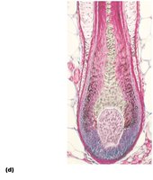
Hair is a flexible strand of dead, keratinized cells. It consists of three concentric layers:
Medulla: Central core.
Cortex: Surrounds the medulla.
Cuticle: Outermost layer.
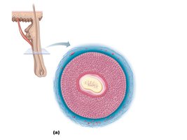
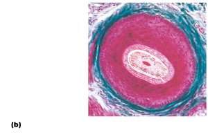
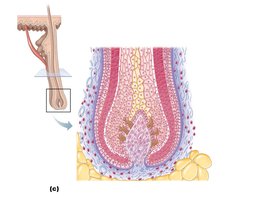
Hair follicles extend from the epidermis into the dermis. The hair bulb is the deep, expanded end of the follicle, surrounded by a root plexus (sensory nerves). The wall of the follicle consists of a peripheral connective tissue sheath, glassy membrane, and epithelial root sheath. The arrector pili muscle, a bundle of smooth muscle, causes hair to stand erect when contracted.
Types and Growth of Hair
Vellus Hairs: Fine body hairs of women and children.
Terminal Hairs: Coarser hair of scalp, axillary, and pubic regions.
Hair Thinning and Baldness: Occur with aging; male pattern baldness is common.
Skin Glands
Sebaceous Glands
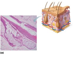
Sebaceous glands are simple alveolar glands that secrete sebum, an oily substance, via holocrine secretion (entire cell breaks up). Sebum collects dirt, softens, and lubricates hair and skin. Most sebaceous glands are associated with hair follicles and are absent from palms and soles.
Sweat Glands
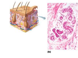
Sweat glands (sudoriferous glands) are widely distributed and produce sweat, a blood filtrate composed of 99% water, salts, and traces of metabolic wastes (urea, ammonia, uric acid). There are two main types:
Eccrine Glands: Most numerous; produce true sweat.
Apocrine Glands: Confined to axillary, anal, and genital areas; produce a special kind of sweat with a musky odor. Modified apocrine glands include ceruminous (ear wax) and mammary (milk) glands.
Burns
Classification and Clinical Relevance
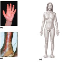
First-Degree Burn: Only the upper epidermis is damaged.
Second-Degree Burn: Upper part of dermis is also damaged; blisters appear; heals with little scarring.
Third-Degree Burn: Consumes the thickness of skin; area appears white, red, or blackened.
The Rule of Nines is used to estimate the extent of burns:
Body Region | Percentage |
|---|---|
Head and Neck | 9% |
Upper Limbs | 18% |
Trunk | 36% |
Lower Limbs | 36% |
Perineum | 1% |
Total | 100% |
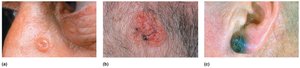
Skin Cancer
Types and Characteristics
Basal Cell Carcinoma: Least malignant and most common; arises from basal cells.
Squamous Cell Carcinoma: Arises from keratinocytes of the stratum spinosum.
Melanoma: Cancer of melanocytes; most dangerous type.
The Skin Throughout Life
Changes with Aging
Skin thins and becomes less elastic from middle to old age.
Shows harmful effects of environmental damage.
Skin inflammations become more common.
