 Back
BackChapter 6: Bones and Bone Structure – Study Guide
Study Guide - Smart Notes
 Tailored notes based on your materials, expanded with key definitions, examples, and context.
Chapter 6: Bones and Bone Structure
Introduction to the Skeletal System
The skeletal system is a fundamental component of human anatomy, providing structure, protection, and support for the body. It consists of bones, cartilages, ligaments, and other connective tissues that stabilize or interconnect the bones.
Structural Support: Bones form the framework for the body.
Mineral and Lipid Storage: Bones store minerals (especially calcium) and lipids (yellow bone marrow).
Blood Cell Production: Red bone marrow produces blood cells.
Protection: Bones protect vital organs (e.g., heart, lungs, brain).
Leverage for Movement: Bones act as levers for muscles, enabling movement.
Classification of Bones by Shape
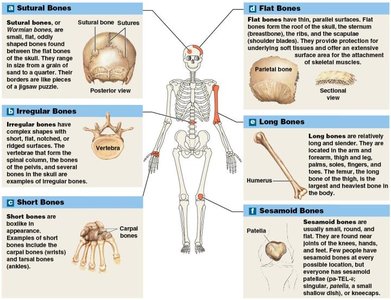
Bones are classified into six broad categories based on their shape, each with distinct structural and functional characteristics.
Sutural Bones: Small, flat, irregular bones found between the flat bones of the skull. Associated with cranial sutures.
Irregular Bones: Complex shapes, such as vertebrae and pelvic bones.
Short Bones: Boxy bones, e.g., carpals (wrist) and tarsals (ankle).
Flat Bones: Thin bones with parallel surfaces, e.g., skull roof, sternum, ribs, scapulae.
Long Bones: Long and slender, found in arms, legs, fingers, toes (e.g., humerus, femur).
Sesamoid Bones: Small, round, flat bones found within tendons near joints (e.g., patella).
Bone Markings
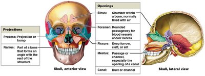
Bones display characteristic surface features known as bone markings, which are essential for muscle attachment, joint formation, and passage of blood vessels and nerves.
Projections: Sites for muscle, tendon, and ligament attachment; also part of articulations (joints).
Openings: Allow passage of blood vessels and nerves.
Depressions: Sites where blood vessels or nerves lie alongside or penetrate bone.
Bone Structure
Bones have distinct structural regions, each with specific functions. The structure varies between long bones and flat bones.
Long Bone Structure:
Diaphysis: Tubular shaft with compact bone surrounding a medullary cavity.
Epiphysis: Ends of the bone, mostly spongy bone.
Metaphysis: Narrow area connecting diaphysis and epiphysis.
Flat Bone Structure: Core of spongy bone between two layers of compact bone.
Bone Tissue and Matrix
Bone tissue is a specialized connective tissue with a dense matrix composed of calcium salts and collagen fibers. Osteocytes are mature bone cells found in lacunae.
Canaliculi: Narrow passageways for nutrient and waste exchange.
Periosteum: Membrane covering bone surfaces, with fibrous and cellular layers.
Bone Matrix:
2/3 weight: Calcium phosphate forms hydroxyapatite crystals with calcium hydroxide .
1/3 weight: Collagen fibers provide flexibility and framework for mineral deposition.
Combination of collagen and hydroxyapatite gives bone strength and resistance to compression.
Bone Cells
Four types of bone cells regulate bone formation, maintenance, and resorption.
Osteogenic Cells: Stem cells that produce osteoblasts; important for fracture repair.
Osteoblasts: Immature cells that synthesize new bone matrix (osteoid) and promote mineralization.
Osteocytes: Mature bone cells in lacunae; maintain bone matrix and assist in repair.
Osteoclasts: Multinucleate cells that resorb bone matrix (osteolysis); derived from macrophage lineage.
Compact Bone
Compact bone is organized into osteons, which are cylindrical structures containing concentric lamellae and a central canal for blood vessels.
Osteon: Basic unit; central canal (Haversian canal) contains blood vessels.
Perforating Canals: (Volkmann’s canals) supply blood to deep osteons and medullary cavity.
Lamellae: Three types: concentric (form osteons), interstitial (fill spaces), circumferential (outer/inner surfaces).
Spongy Bone
Spongy bone lacks osteons and consists of trabeculae, which support red bone marrow. Blood vessels in red marrow supply nutrients to osteocytes.
Red Bone Marrow: Site of blood cell formation.
Yellow Bone Marrow: Energy reserve (adipose tissue); found in medullary cavities of long bones in adults.
Bone Membranes
Bones are covered and lined by specialized membranes that play roles in growth, repair, and remodeling.
Periosteum: Covers outer surfaces; isolates bone, provides routes for vessels/nerves, participates in growth/repair.
Endosteum: Lines inner surfaces; active during growth, repair, and remodeling.
Bone Formation and Growth
Bone formation (ossification) occurs via two processes: endochondral and intramembranous ossification. Calcification is the deposition of calcium salts during ossification.
Endochondral Ossification: Bone replaces cartilage; occurs mostly in long bones during fetal development.
Intramembranous Ossification: Bone develops directly from mesenchymal tissue; occurs mostly in flat bones.
Endochondral Ossification Steps
Chondrocytes enlarge at the primary ossification center; matrix calcifies and cells die.
Blood vessels grow around cartilage; perichondrium cells become osteoblasts.
Blood vessels penetrate; fibroblasts become osteoblasts, forming spongy bone.
Spongy bone remodels, creating medullary cavity; shaft thickens.
Blood vessels and osteoblasts migrate into epiphyses, forming secondary ossification centers.
Epiphyses fill with spongy bone; epiphyseal plate separates epiphysis and diaphysis.
At puberty, ossification overtakes cartilage production, leading to epiphyseal closure and formation of epiphyseal line.
Intramembranous Ossification Steps
Mesenchymal cells differentiate into osteoblasts, secrete osteoid, which calcifies.
Osteoblasts surrounded by matrix become osteocytes; bone grows in spicules.
Blood vessels grow between spicules, accelerating growth.
Spongy bone forms via continued deposition.
Remodeling produces compact bone; periosteum forms.
Bone Vessels and Innervation
Bones are highly vascularized and innervated, supporting their metabolic activity and sensitivity to injury.
Nutrient Artery and Vein: Supply diaphysis.
Metaphyseal Vessels: Supply epiphyseal cartilage.
Periosteal Vessels: Supply superficial osteons and secondary ossification centers.
Lymphatic Vessels and Sensory Nerves: Present in periosteum, endosteum, medullary cavity, and epiphyses.
Bone Remodeling
Bone remodeling is a continuous process involving osteocytes, osteoblasts, and osteoclasts, allowing bones to adapt to stress and repair damage.
Osteoclasts: Remove bone matrix.
Osteoblasts: Add bone matrix.
Balance: Determines bone strength; exercise increases bone strength.
Calcium Homeostasis
The skeleton is the primary reservoir for calcium, which is essential for physiological processes. Blood calcium levels are tightly regulated by hormones.
Parathyroid Hormone (PTH): Increases blood calcium by stimulating osteoclasts, increasing intestinal absorption, and decreasing kidney excretion.
Calcitonin: Decreases blood calcium by inhibiting osteoclasts, increasing kidney excretion, and reducing intestinal absorption.
Fractures
Fractures are breaks in bones caused by physical stress. They are classified as open (compound) or closed (simple) based on whether the bone projects through the skin.
Open Fracture: Bone projects through the skin.
Closed Fracture: Bone remains internal.
