 Back
Chapter 6: Bones and Bone Tissue – Comprehensive Study Guide
Study Guide - Smart Notes
 Tailored notes based on your materials, expanded with key definitions, examples, and context.
Functions of the Skeletal System
Overview of Skeletal Functions
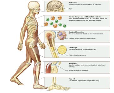
The skeletal system performs several essential functions that are critical for maintaining homeostasis and supporting life. These functions include protection, mineral storage, acid-base balance, blood cell formation, fat storage, movement, and support.
Protection: Bones surround and protect vital organs such as the brain, heart, and lungs.
Mineral Storage and Acid-Base Homeostasis: Bones act as reservoirs for minerals like calcium (Ca2+) and phosphate (PO43−), which are essential for electrolyte and acid-base balance.
Blood Cell Formation: Red bone marrow is the site of hematopoiesis, the process of forming blood cells.
Fat Storage: Yellow bone marrow stores triglycerides, serving as an energy reserve.
Movement: Bones serve as levers for muscles, facilitating movement at joints.
Support: The skeleton provides the structural framework that supports the body’s weight.
Classification of Bones by Shape
Types of Bones
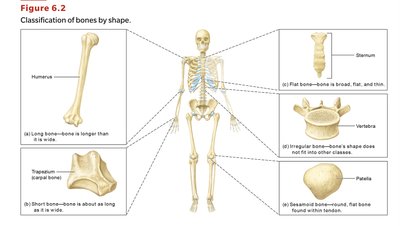
Bones are classified according to their shapes, which relate to their functions and locations in the body.
Long Bones: Longer than they are wide (e.g., humerus, femur, phalanges).
Short Bones: Roughly cube-shaped, about as long as they are wide (e.g., carpals, tarsals).
Flat Bones: Thin, broad, and flat (e.g., sternum, ribs, skull bones, pelvis).
Irregular Bones: Complex shapes that do not fit other categories (e.g., vertebrae).
Sesamoid Bones: Small, oval bones embedded within tendons (e.g., patella).
Gross Anatomy of Bones
Long Bone Structure
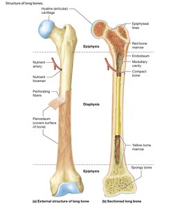
Long bones have a unique structure that supports their function in movement and weight-bearing.
Diaphysis: The shaft or central part of a long bone.
Epiphyses: The enlarged ends of the bone, which articulate with other bones.
Articular Cartilage: Hyaline cartilage covering the epiphyses to reduce friction at joints.
Medullary Cavity: The hollow space within the diaphysis containing bone marrow.
Compact Bone: Dense outer layer that provides strength and resists compression.
Spongy Bone: Internal network of trabeculae that absorbs shock and houses marrow.
Periosteum: Dense connective tissue membrane covering the bone’s outer surface.
Perforating Fibers: Collagen fibers anchoring the periosteum to the bone matrix.
Endosteum: Thin membrane lining the internal bone surfaces and cavities.
Epiphyseal Lines: Remnants of the growth plate in adult bones.
Structure of Short, Flat, Irregular, and Sesamoid Bones
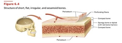
These bones lack a medullary cavity and have a sandwich-like structure.
Sandwich Structure: Two layers of compact bone surround a middle layer of spongy bone (diploë in flat bones).
Sinuses: Air-filled spaces in some skull bones that reduce weight.
Microscopic Structure and Bone Matrix
Importance of Bone Matrices
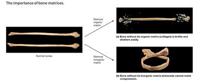
The bone matrix is composed of inorganic and organic components, each contributing to bone’s physical properties.
Inorganic Matrix (65%): Mainly hydroxyapatite crystals [], providing hardness and resistance to compression.
Organic Matrix (35%): Primarily collagen fibers, giving bone flexibility and tensile strength.
Deficiency Effects: Without collagen, bones are brittle; without minerals, bones are flexible and cannot resist compression.
Cellular Components of Bone Tissue
Types of Bone Cells
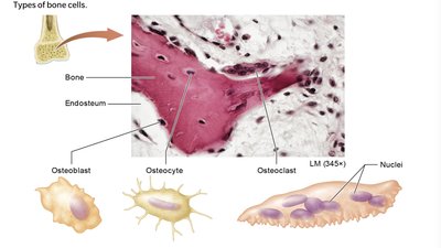
Bone tissue is dynamic, maintained by three main cell types:
Osteoblasts: Immature, bone-building cells found on bone surfaces; responsible for bone deposition.
Osteocytes: Mature bone cells trapped in lacunae; maintain bone matrix and sense mechanical stress.
Osteoclasts: Large, multinucleated cells responsible for bone resorption; break down bone matrix using acids and enzymes.
Functions of Osteoblasts and Osteocytes
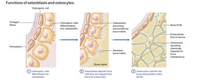
Osteoblasts: Deposit bone matrix until they are trapped and become osteocytes.
Osteocytes: Maintain the extracellular matrix and communicate with other bone cells to regulate bone health.
Function of Osteoclasts
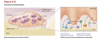
Osteoclasts: Secrete hydrogen ions and enzymes to dissolve bone matrix, releasing minerals into the blood.
Resorption: Critical for bone remodeling and calcium homeostasis.
Histology of Bone Tissue
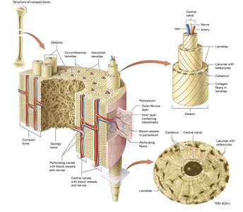
Structure of Compact Bone
Compact bone is organized into osteons, which are cylindrical structures that provide strength and house blood vessels and nerves.
Osteon (Haversian System): The basic unit, consisting of concentric lamellae around a central canal.
Central Canal: Contains blood vessels and nerves.
Perforating Canals: Connect central canals transversely.
Lacunae: Small spaces housing osteocytes between lamellae.
Canaliculi: Tiny channels connecting lacunae for nutrient and signal exchange.
Structure of Spongy Bone
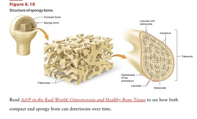
Spongy bone consists of a network of trabeculae, which provide structural support and house bone marrow.
Trabeculae: Lattice-like bony struts covered by endosteum.
Lacunae and Canaliculi: House osteocytes and allow for nutrient diffusion.
Bone Formation (Ossification)
Intramembranous Ossification
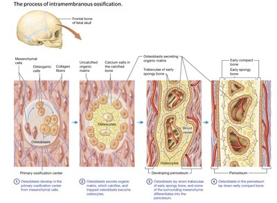
This process forms flat bones (e.g., skull, clavicles) from a membrane of embryonic connective tissue (mesenchyme).
Primary Ossification Center: Site where osteoblasts begin secreting matrix.
Sequence: Mesenchymal cells differentiate into osteoblasts → osteoblasts secrete matrix → matrix calcifies → osteocytes form → spongy bone forms → periosteum develops → compact bone forms externally.
Endochondral Ossification
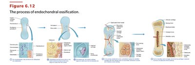
This process forms most bones below the head (except clavicles) from a hyaline cartilage model.
Sequence: Perichondrium converts to periosteum → osteoblasts build bone collar → cartilage calcifies and dies → blood vessels and osteoblasts invade → spongy bone forms → medullary cavity develops → epiphyses ossify.
Cartilage Remnants: Hyaline cartilage remains at the epiphyseal plates (growth plates) and articular surfaces.
Bone Growth
Longitudinal Growth (Length)
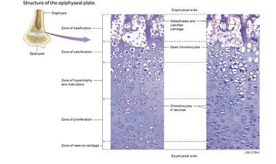
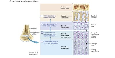
Occurs at the epiphyseal plate through the division and maturation of chondrocytes.
Zones (from epiphysis to diaphysis):
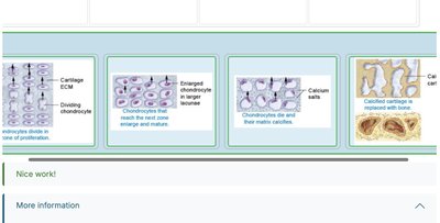
Zone of Proliferation: Chondrocytes divide.
Zone of Hypertrophy & Maturation: Chondrocytes enlarge.
Zone of Calcification: Chondrocytes die, matrix calcifies.
Zone of Ossification: Osteoblasts replace cartilage with bone.
Epiphyseal Plate Closure: Occurs between ages 13–21, forming the epiphyseal line and ending longitudinal growth.
Appositional Growth (Width)
Bones grow in width throughout life by adding new bone to the surface beneath the periosteum while osteoclasts resorb bone from the medullary cavity.
Result: Bone thickens and strengthens without becoming too heavy.
Bone Remodeling and Calcium Homeostasis
Bone Remodeling
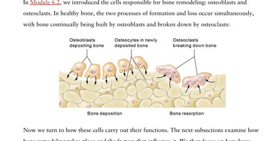
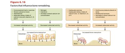
Bone remodeling is a continuous process involving bone deposition by osteoblasts and bone resorption by osteoclasts. It maintains bone strength and mineral homeostasis.
Deposition: Osteoblasts build new bone matrix.
Resorption: Osteoclasts break down old bone matrix.
Mechanical Stress: Weight-bearing activity increases deposition; lack of stress increases resorption.
Calcium Ion Homeostasis
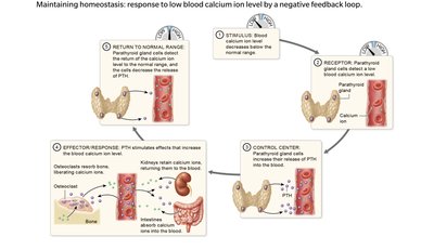
Calcium levels are tightly regulated by hormones through a negative feedback loop.
Low Blood Calcium: Detected by parathyroid glands, which release parathyroid hormone (PTH).
PTH Effects: Stimulates osteoclasts to release calcium from bone, increases calcium reabsorption in kidneys, and enhances intestinal absorption (with vitamin D).
High Blood Calcium: Triggers calcitonin release from the thyroid gland, which inhibits osteoclasts and increases bone deposition.
Fracture Repair
Steps of Fracture Repair
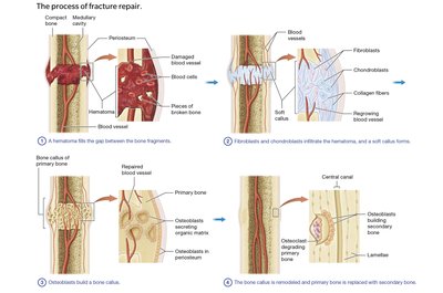
Bone repair after a fracture occurs in four main steps:
Fracture Hematoma Formation: Blood vessels rupture, forming a clot.
Soft Callus Formation: Fibroblasts and chondroblasts build a flexible bridge of collagen and cartilage.
Bone Callus Formation: Osteoblasts lay down primary bone, forming a hard callus.
Remodeling: Osteoclasts and osteoblasts remodel the callus into mature secondary bone.
Types of Bone Fractures
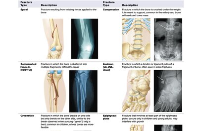
Fractures are classified by their pattern and the forces involved. Common types include spiral, compression, comminuted, avulsion, greenstick, and epiphyseal plate fractures.
Spiral: Twisting force causes a spiral break.
Compression: Bone is crushed, often in vertebrae.
Comminuted: Bone shatters into multiple fragments.
Avulsion: Tendon or ligament pulls off a bone fragment.
Greenstick: Bone bends and cracks, common in children.
Epiphyseal Plate: Involves the growth plate, may affect growth in children.
Summary Table: Bone Matrix Components and Effects of Deficiency
Component | Main Function | Deficiency Effect |
|---|---|---|
Inorganic Matrix (Hydroxyapatite) | Hardness, compression resistance | Bone becomes flexible, cannot resist compression |
Organic Matrix (Collagen) | Tensile strength, flexibility | Bone becomes brittle, shatters easily |
Key Equations
Hydroxyapatite Formula:
Additional info:
Vitamin D is essential for calcium absorption; deficiency leads to rickets in children.
Bone remodeling adapts the skeleton to mechanical stress and repairs micro-damage.
Hormones such as growth hormone, estrogen, and testosterone regulate bone growth and remodeling.
