 Back
BackChapter 6: Bones & Skeletal Tissues – Study Guide
Study Guide - Smart Notes
 Tailored notes based on your materials, expanded with key definitions, examples, and context.
Bones & Skeletal Tissues
Overview
Bones are complex organs composed of several tissue types, including bone tissue, collagen, epithelial tissue, nerve tissue, and muscular tissue. The skeletal system is formed by various types of cartilage and bone, each with distinct structural and functional properties.
Types of Cartilage in the Skeleton
Hyaline, Elastic, and Fibrocartilage
Cartilage is a flexible connective tissue that plays a crucial role in the formation and function of the skeleton.
Hyaline Cartilage: Appears glassy, provides support with flexibility and resilience. Most abundant; found in articular, costal, respiratory, and nasal cartilages.
Elastic Cartilage: Contains more elastic fibers, withstands repeated bending. Found in the external ear and epiglottis.
Fibrocartilage: Highly compressible, great tensile strength. Found in menisci of the knee and intervertebral discs.
Cartilage Growth: Cartilage grows by appositional (external) and interstitial (internal) mechanisms, accommodating mitosis and rapid skeletal development.
Functions of Bone
Major Functions
Bones perform several essential functions:
Support: Framework for the body
Protection: Shields vital organs
Anchorage: Attachment for muscles, enabling movement
Mineral Storage: Stores calcium and phosphate
Blood Cell Formation: Occurs in red marrow
Fat Storage: Yellow marrow stores fat
Hormone Production: Osteocalcin regulates bone formation
Classification of Bones
By Location and Shape
Bones are classified as axial or appendicular, and by shape: long, short (including sesamoid and sutural), flat, and irregular.
Gross Structure of Bones
Compact and Spongy Bone
Bones consist of two main types of tissue: compact and spongy bone.
Compact Bone: Dense, forms the outer layer, composed of osteons.
Spongy Bone: Lattice-like network of trabeculae, found inside bones.
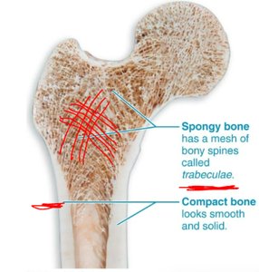
Structure of Long Bones: Includes diaphysis, epiphysis, metaphysis, medullary cavity, and articular cartilage.
Hematopoietic Tissue: Red marrow in spongy bone produces blood cells.
Microscopic Anatomy of Bone
Bone Cells
Bone tissue contains several cell types:
Osteoprogenitor Cells: Stem cells that differentiate into osteoblasts.
Osteoblasts: Bone-building cells, secrete new bone matrix.
Osteocytes: Mature bone cells, maintain matrix, act as mechanosensors.
Osteoclasts: Bone-resorbing cells, degrade bone tissue via osteolytic enzymes.
Microscopic Anatomy of Compact Bone: Osteons (Haversian systems) are the structural units, consisting of lamellae, osteocytes, central (Haversian) canal, perforating (Volkmann's) canal, interstitial and circumferential lamellae.
Microscopic Anatomy of Spongy Bone: Composed of trabeculae.
Chemical Composition of Bone
Organic and Inorganic Components
Organic: Collagen fibers (osteoid), provides tensile strength.
Inorganic: Hydroxyapatites (mineral salts, mainly calcium phosphate), provides hardness and resistance to compression.
Bone Development (Ossification)
Intramembranous and Endochondral Ossification
Bone formation occurs via two processes:
Endochondral Ossification: Uses hyaline cartilage as a model; most bones form this way. Steps include bone collar formation, cartilage calcification, periosteal bud invasion, diaphysis elongation, and epiphysis ossification.
Intramembranous Ossification: Bone develops directly within fibrous connective tissue; forms flat bones, mandible, clavicle.
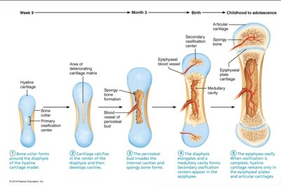
Postnatal Bone Growth
Growth in Length and Width
Long bones grow in length via the epiphyseal plate, which consists of several zones:
Proliferation Zone: Cartilage cells undergo mitosis.
Hypertrophic Zone: Older cartilage cells enlarge.
Calcification Zone: Matrix calcifies, cartilage cells die.
Ossification Zone: New bone forms.
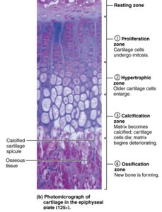
Growth in width (appositional growth) occurs in all bones. Hormones such as growth hormone, thyroid hormone, and estrogen regulate bone growth.
Bone Remodeling
Bone Deposition and Resorption
Bone remodeling is a continuous process involving bone deposition (by osteoblasts) and resorption (by osteoclasts). Remodeling maintains calcium homeostasis and bone strength.
Bone Resorption: Releases minerals, remodels bone.
Bone Deposition: Stores minerals, forms new bone.
Hormonal Control of Remodeling
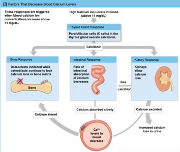
Calcium levels in blood are regulated by hormones:
Parathyroid Hormone (PTH): Increases blood calcium by stimulating osteoclasts, increasing intestinal absorption, and conserving calcium in kidneys.
Calcitonin: Decreases blood calcium by inhibiting osteoclasts, reducing intestinal absorption, and increasing calcium excretion in kidneys.
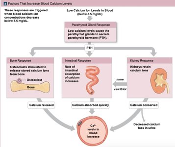
Response to Mechanical Stress
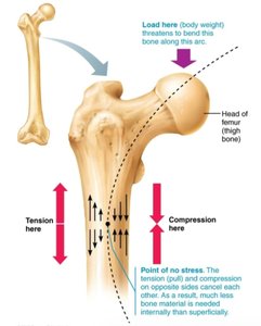
According to Wolff's Law, bones remodel in response to mechanical and gravitational forces. Areas subjected to stress become stronger.
Bone Repair
Fracture Classification and Repair
Fractures are classified by their nature (closed/simple, open/compound, displaced, nondisplaced, compression, spiral, epiphyseal, comminuted, greenstick, depressed). Bone repair involves four steps:
Hematoma formation
Fibrocartilaginous callus formation
Bony callus formation
Bone remodeling
Bone Disorders
Osteomalacia, Rickets, Osteoporosis, Paget's Disease
Osteomalacia and Rickets: Softening of bones due to vitamin D deficiency.
Osteoporosis: Reduced bone mass, increased fracture risk.
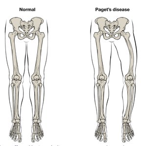
Paget's Disease: Abnormal bone remodeling, resulting in porous and curved bones.
Summary Table: Cartilage vs. Bone (Inferred from Table 6.1)
Feature | Cartilage | Bone |
|---|---|---|
Matrix | Flexible | Hard |
Cell Type | Chondrocytes | Osteocytes |
Growth | Appositional & Interstitial | Appositional only |
Blood Supply | Avascular | Vascular |
Function | Support, flexibility | Support, protection, mineral storage |
Additional info: Table inferred based on lecture notes and typical textbook content.
