 Back
BackCompartmentation: Cells and Tissues – Structured Study Notes
Study Guide - Smart Notes
 Tailored notes based on your materials, expanded with key definitions, examples, and context.
Compartmentation: Cells and Tissues
Introduction to Physiology and Compartmentation
Physiology is the study of normal organismal functioning and its component parts. The cell is the basic functional unit of living organisms, capable of carrying out all processes of life. However, no single cell can perform all the processes required by the mature human body; thus, cells assemble into tissues, which work together to achieve common purposes.
Functional Compartments of the Body
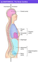
The human body is organized into several major anatomical and functional compartments, each with specific boundaries and contents.
Major Body Cavities: Cranial (skull), Thoracic (ribs & diaphragm), and Abdominopelvic (peritoneum surrounds organs; kidneys are outside this cavity).
Fluid-filled Compartments: Circulatory system, eyes, cerebrospinal fluid (CSF), pleural and pericardial sacs.
Lumens: The interior of hollow organs (e.g., heart, lungs, blood vessels, intestines) is called the lumen, which may be filled with air or fluid. In some organs, the lumen is an extension of the external environment (e.g., food in the intestines is technically outside the body until absorbed).
The Body’s Fluid Compartments
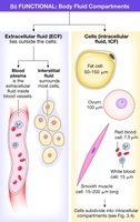
Body fluids are separated into two main compartments by cell membranes:
Intracellular Fluid (ICF): Fluid inside cells.
Extracellular Fluid (ECF): Fluid outside cells, further divided into plasma (fluid portion of blood) and interstitial fluid (surrounds most cells).
Cell Membranes
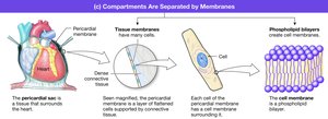
Structure and Function of Cell Membranes
The cell membrane (also called plasma membrane or plasmalemma) is a thin lipid bilayer that separates the cell’s interior from the external environment. It is composed mainly of phospholipids and proteins, with the ratio varying by cell type. Metabolically active cells have higher protein content in their membranes.
Functions:
Physical isolation (separates ECF from ICF)
Regulation of exchange (controls entry/exit of ions, nutrients, waste)
Communication (contains proteins for signaling and recognition)
Structural support (anchors cell to cytoskeleton and extracellular matrix)
Fluid Mosaic Model
The fluid mosaic model describes the cell membrane as a dynamic, three-dimensional structure composed of phospholipids, proteins, glycoproteins, glycolipids, and cholesterol. These components are not fixed and can move laterally within the membrane.
Membrane Lipids and Proteins
Membrane lipids create a hydrophobic barrier that prevents water-soluble (hydrophilic) molecules from crossing without assistance. Membrane proteins are classified as:
Integral proteins: Tightly bound, including transmembrane and lipid-anchored proteins.
Peripheral proteins: Loosely bound, often enzymes or structural proteins.

Membrane Carbohydrates
Carbohydrates attach to both lipids (glycolipids) and proteins (glycoproteins) on the external surface of the cell, forming the glycocalyx. This layer is important for protection and immune response (e.g., ABO blood groups).
Membrane Composition Table
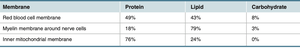
The composition of membranes varies by cell type. The following table summarizes the protein, lipid, and carbohydrate content of selected membranes:
Membrane | Protein | Lipid | Carbohydrate |
|---|---|---|---|
Red blood cell membrane | 49% | 43% | 8% |
Myelin membrane (nerve cells) | 18% | 79% | 3% |
Inner mitochondrial membrane | 76% | 24% | 0% |
Intracellular Compartments
Major Components of the Cell
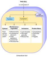
Cells are divided into compartments, each with specialized functions:
Cell Membrane
Nucleus: Contains genetic material and directs cell functions.
Cytoplasm: Includes cytosol (ICF), inclusions (e.g., nutrients, ribosomes), protein fibers (cytoskeleton), and organelles (membrane-bound structures).
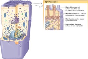
Cytoplasmic Protein Fibers and Cytoskeleton
The cytoskeleton is a dynamic network of protein fibers that provides structural support, facilitates movement, and organizes the cell internally. There are three main types of protein fibers:
Diameter | Type of Protein | Functions |
|---|---|---|
Microfilaments (7 nm) | Actin | Cell shape, muscle contraction |
Intermediate Filaments (10 nm) | Keratin, neurofilament | Structural support, barrier function |
Microtubules (25 nm) | Tubulin | Movement of cilia/flagella, intracellular transport |
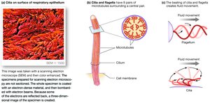
Cilia and Flagella
Microtubules form cilia and flagella, which are structures for cellular movement. Both have a 9+2 arrangement of microtubules and use the motor protein dynein. Cilia beat rhythmically to move fluids, while flagella (e.g., in sperm) move in a wavelike fashion.
Cytoskeleton Functions
Maintains cell shape
Organizes internal cell structure
Facilitates intracellular transport
Enables cell movement and division
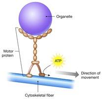
Motor Proteins
Motor proteins use ATP to move along cytoskeletal fibers, transporting organelles and vesicles. Major types include myosins (muscle contraction), kinesins, and dyneins (vesicle and cilia/flagella movement).
Organelles and Their Functions
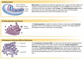
Mitochondria: Site of ATP production; contains its own DNA.
Endoplasmic Reticulum (ER): Rough ER synthesizes proteins; smooth ER synthesizes lipids, fatty acids, and steroids.
Golgi Apparatus: Modifies, sorts, and packages proteins and lipids for secretion or delivery to other organelles.
Lysosomes: Contain hydrolytic enzymes for intracellular digestion and autophagy.
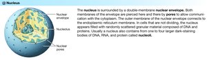
Nucleus
The nucleus is the cell’s control center, containing chromatin (DNA and proteins) and directing all cellular functions. The nuclear envelope is a double membrane with pores for communication with the cytoplasm. The nucleolus is the site of ribosomal RNA synthesis.
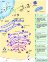
Protein Synthesis and Compartmentation
Protein synthesis, modification, and packaging occur in different cell compartments, demonstrating the importance of compartmentation for cellular function.
Tissues of the Body (Histology)
Four Basic Tissue Types
Tissues are groups of cells held together by cell junctions. The four primary tissue types are:
Epithelial: Protection, regulation of exchange between internal and external environments.
Connective: Structural support, physical barrier, defense against invaders.
Muscle: Contraction, force, and movement (cardiac, smooth, skeletal).
Neural: Communication via electrical and chemical signals (neurons and glial cells).
Cell Junctions
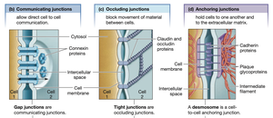
Three main types of cell junctions connect cells within tissues:
Communicating (gap) junctions: Allow direct cell-to-cell communication.
Occluding (tight) junctions: Block movement of materials between cells.
Anchoring junctions: Hold cells to each other and the extracellular matrix.
Epithelial Tissue
Epithelial tissue forms layers around surfaces and can be simple (one cell thick) or stratified (multiple layers). The apical membrane faces the lumen, while the basolateral membrane faces the extracellular fluid. All substances entering or leaving the body must cross an epithelium.
Functional Categories of Epithelia
Exchange Epithelium: Thin, flattened cells for gas exchange (e.g., blood vessels, lungs; called endothelium in heart and vessels).
Transporting Epithelium: Cuboidal or columnar cells with specialized membranes for selective transport.
Ciliated Epithelium: Cilia move fluids and particles (e.g., respiratory tract, female reproductive tract).
Protective Epithelium: Multiple layers to prevent exchange and protect against stress.
Secretory Epithelium: Produce and secrete substances; may form glands (exocrine and endocrine).
Connective, Muscle, and Neural Tissues
Connective Tissue: Includes blood, bone, cartilage, and support tissues for organs and skin. Provides structure and defense.
Muscle Tissue: Contracts to produce movement; types include cardiac, smooth, and skeletal muscle.
Neural Tissue: Neurons transmit signals; glial cells support neurons. Both are excitable tissues capable of generating action potentials.
Organs and Integration of Tissues
Organs are structures composed of two or more tissue types that work together to perform specific functions. The skin is an example of an organ that integrates all four tissue types (epithelial, connective, muscle, neural) into a functional whole.
Apoptosis
Apoptosis is programmed cell death, a normal process in which cells shrink and fragment into membrane-bound blebs that are removed by neighboring or immune cells. This process is essential for development and tissue homeostasis.
