 Back
BackComprehensive Study Notes: Anatomy and Physiology of the Respiratory System
Study Guide - Smart Notes
 Tailored notes based on your materials, expanded with key definitions, examples, and context.
Respiratory System
Functional Anatomy of the Respiratory System
The respiratory system is responsible for gas exchange between the atmosphere and body cells, facilitating oxygen intake and carbon dioxide removal. It is divided into upper and lower respiratory tracts, each with specialized structures for air conduction and filtration.
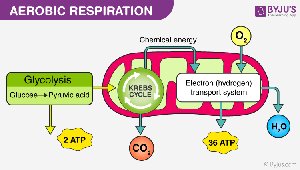
Gas Exchange: Involves ventilation, external respiration (air/lungs), gas transport, internal respiration (blood/cells), and cellular respiration (O2/CO2).
Why We Breathe: Oxygen is essential for cellular energy production via the electron transport chain, producing CO2 and ATP.
Organs of the Respiratory System
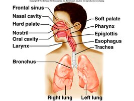
The respiratory system consists of several organs organized into the upper and lower respiratory tracts.
Upper Respiratory Tract: Nose, nasal cavity, sinuses, pharynx
Lower Respiratory Tract: Larynx, trachea, bronchial tree, lungs
Upper Respiratory Tract Anatomy
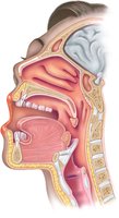
The upper respiratory tract includes structures that filter, warm, and humidify incoming air.
Nasal Conchae: Superior, middle, and inferior conchae increase surface area for air filtration.
Sinuses: Air-filled spaces within maxillary, frontal, ethmoid, and sphenoid bones.
Pharynx: Passageway for air and food, divided into nasopharynx, oropharynx, and laryngopharynx.
Respiratory Epithelium
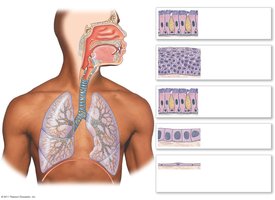
The lining of the respiratory tract varies by region, providing protection and facilitating mucous movement.
Pseudostratified Ciliated Columnar Epithelium: Lines most of the upper respiratory tract, moves mucous and trapped particles.
Stratified Squamous Epithelium: Found in areas subject to abrasion, such as the oropharynx.
Simple Cuboidal and Simple Squamous Epithelium: Present in smaller airways and alveoli, respectively.
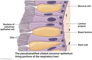
Mucous and Cilia in the Respiratory Tract
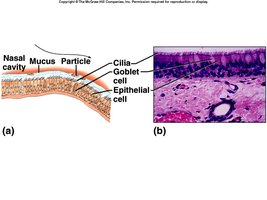
Mucous traps particles, and cilia move them toward the pharynx for removal, protecting the lower airways.
Goblet Cells: Produce mucous.
Cilia: Propel mucous and debris out of the respiratory tract.
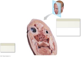
Respiratory Sinuses
Sinuses are air-filled spaces that lighten the skull and contribute to voice resonance.
Maxillary, Frontal, Ethmoid, Sphenoid Sinuses: Each sinus is located within its respective bone.
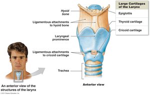
Larynx and Vocal Cords
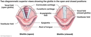
The larynx is a passageway for air and houses the vocal cords, which produce sound.
Vocal Cords: True vocal cords are responsible for vocal sounds; pitch and intensity are controlled by tension and air force.
Trachea
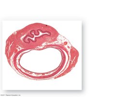
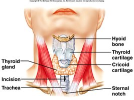
The trachea transports air between the larynx and bronchi, supported by cartilage rings.
Cartilage: Maintains airway patency.
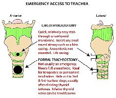
Clinical Applications: Tracheostomy and cricothyroidotomy allow air to bypass obstructions.
Bronchial Tree
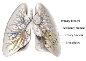
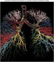
The bronchial tree consists of branched airways leading from the trachea to the alveoli, facilitating air distribution.
Primary Bronchi: Right and left bronchi branch from the trachea.
Secondary and Tertiary Bronchi: Further subdivisions within the lungs.
Bronchioles: Smallest conducting airways, ending in terminal and respiratory bronchioles.
Alveolar Ducts, Sacs, and Alveoli: Sites of gas exchange.
Alveoli and Gas Exchange
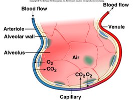
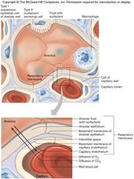
Alveoli are tiny air sacs where oxygen and carbon dioxide are exchanged between air and blood.
Respiratory Membrane: Composed of alveolar and capillary walls, facilitating diffusion.
Gas Exchange: Oxygen diffuses from alveolus to capillary; carbon dioxide diffuses from capillary to alveolus.
Lungs
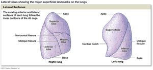
The lungs are soft, spongy organs located in the thoracic cavity, divided into lobes.
Right Lung: Three lobes (superior, middle, inferior).
Left Lung: Two lobes (superior, inferior).
Respiratory Physiology
Breathing Mechanism
Breathing, or ventilation, is the movement of air into and out of the lungs, driven by pressure changes.
Inspiration: Inhaling; intra-alveolar pressure decreases as thoracic cavity enlarges, allowing air to enter.
Expiration: Exhaling; elastic recoil of lung tissues and abdominal organs expels air.
Boyle’s Law
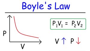
Boyle’s Law states that pressure is inversely proportional to volume in a closed system, explaining how lung volume changes affect air pressure.
Equation:
Application: As lung volume increases, pressure decreases, drawing air in.
Lung Volumes and Capacities
Lung volumes and capacities are measurements of air movement during breathing.
Tidal Volume (TV): ~500 mL; air entering and leaving lungs per breath.
Inspiratory Reserve Volume (IRV): ~3000 mL; additional air inhaled.
Expiratory Reserve Volume (ERV): ~1100 mL; additional air exhaled.
Residual Volume (RV): ~1200 mL; air remaining after maximal exhalation.
Vital Capacity (VC):
Total Lung Capacity (TLC):
Nonrespiratory Air Movements
Movements such as coughing, sneezing, laughing, crying, hiccupping, yawning, and speech are nonrespiratory air movements that clear airways or produce sound.
Control of Breathing
Breathing is regulated by respiratory centers in the brainstem, including the medulla oblongata and pons.
Dorsal Respiratory Group (DRG): Maintains constant breathing rhythm.
Ventral Respiratory Group (VRG): Controls forced breathing.
Pontine Respiratory Group: Modifies depth and rate of breathing.
Factors Affecting Breathing
Breathing is influenced by blood oxygen and carbon dioxide levels, pH, and stretch receptors in the lungs.
Peripheral Chemoreceptors: Located in carotid and aortic bodies, respond to decreased blood oxygen.
Stretch Receptors: Prevent overinflation of the lungs.
Alveolar Gas Exchange
Gas exchange occurs due to differences in partial pressure between alveolar air and blood.
Oxygen Transport: 98% bound to hemoglobin, 2% dissolved in plasma.
Carbon Dioxide Transport: 70% as bicarbonate ion, 23% bound to hemoglobin, 7% dissolved in plasma.
Oxygen Release: Increases with higher PCO2 and lower pH.
Clinical Applications
Clinical interventions such as tracheostomy and cricothyroidotomy are used to bypass airway obstructions and restore breathing.
Additional info: Expanded explanations and context were added to ensure completeness and academic quality for college-level study.
