 Back
Comprehensive Study Notes: Blood (Chapter 10, Human Anatomy & Physiology II)
Study Guide - Smart Notes
 Tailored notes based on your materials, expanded with key definitions, examples, and context.
Blood: Overview and Functions
Introduction to Blood
Blood is a specialized connective tissue that plays a vital role in transporting substances throughout the body. It circulates through blood vessels, delivering oxygen, nutrients, hormones, and removing wastes. Blood also helps regulate body temperature and maintain homeostasis.
Transports: Oxygen, nutrients, wastes, hormones, and body heat.
Volume: Approximately 8% of body weight (5–6 liters in adults).
Physical Characteristics: Sticky, opaque, more viscous than water, metallic taste, pH 7.35–7.45, temperature slightly higher than body temperature.
Components of Blood
Main Components
Blood consists of two main components: formed elements (cellular components) and plasma (nonliving fluid matrix).
Formed Elements: Erythrocytes (red blood cells), leukocytes (white blood cells), and platelets.
Plasma: Straw-colored fluid, 90% water, contains dissolved substances (nutrients, salts, respiratory gases, hormones, plasma proteins, waste products).
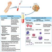
Separation of Blood Components
Plasma: 55% of blood, rises to the top when centrifuged.
Erythrocytes: 45% of blood (hematocrit), sink to the bottom.
Buffy Coat: Less than 1%, contains leukocytes and platelets, appears as a thin whitish layer between plasma and erythrocytes.
Plasma
Composition and Functions
Plasma is the liquid matrix of blood, primarily composed of water and various solutes.
Water (90%): Acts as a solvent for carrying other substances.
Plasma Proteins: Most abundant solutes, mainly produced by the liver.
Key Plasma Proteins:
Albumin: Maintains osmotic pressure and acts as a blood buffer.
Clotting Proteins: Essential for blood clotting (e.g., fibrinogen, prothrombin).
Antibodies: Produced by lymphocytes, provide immune defense.
Other Solutes: Nutrients, electrolytes, respiratory gases, hormones, waste products.
Formed Elements
Overview
The formed elements of blood include erythrocytes, leukocytes, and platelets. Each type has distinct structure and function.
Erythrocytes (RBCs): Transport oxygen and a small amount of carbon dioxide.
Leukocytes (WBCs): Defend the body against infection and disease.
Platelets: Cell fragments essential for blood clotting.
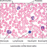
Erythrocytes (Red Blood Cells)
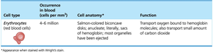
Erythrocytes are specialized for oxygen transport. They are anucleate, lack most organelles, and are packed with hemoglobin.
Shape: Biconcave discs, increasing surface area for gas exchange.
Count: ~5 million per mm3 of blood.
Hemoglobin: Iron-containing protein that binds oxygen; each molecule can carry 4 oxygen molecules.
ATP Production: Anaerobic, so RBCs do not use the oxygen they transport.
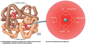
Homeostatic Imbalances of RBCs
Anemia: Decreased oxygen-carrying capacity due to low RBC count or abnormal hemoglobin.
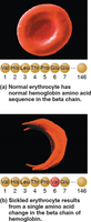
Sickle Cell Anemia: Genetic disorder causing abnormal hemoglobin shape, leading to sickled RBCs and impaired oxygen delivery.
Polycythemia: Excessive or abnormal increase in RBCs, increasing blood viscosity and slowing flow. Causes include bone marrow cancer or adaptation to high altitude.
Leukocytes (White Blood Cells)
Leukocytes are complete cells with nuclei and organelles. They defend the body against pathogens and can move out of blood vessels (diapedesis) to sites of infection.
Count: 4,800–10,800 per mm3 of blood.
Movement: Amoeboid motion, positive chemotaxis (move toward chemical signals from damaged tissue).
Types of Leukocytes
Granulocytes: Contain granules in cytoplasm; lobed nuclei. Includes neutrophils, eosinophils, basophils.
Agranulocytes: Lack visible granules; nuclei are spherical, oval, or kidney-shaped. Includes lymphocytes and monocytes.
Granulocytes
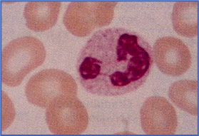
Neutrophils: Most abundant WBC, multilobed nucleus, phagocytic, increase during infection.
Eosinophils: Kill parasitic worms, involved in allergy responses.
Basophils: Rarest WBC, contain histamine (inflammatory response) and heparin (anticoagulant).
Agranulocytes
Lymphocytes: Large, dark nucleus, reside in lymphatic tissues, play a role in immune response.
Monocytes: Largest WBC, U- or kidney-shaped nucleus, become macrophages in tissues, fight chronic infection.
Platelets
Origin: Fragments of megakaryocytes.
Function: Essential for blood clotting.
Count: ~300,000 per mm3 of blood.
Hematopoiesis (Blood Cell Formation)
Process and Sites
Hematopoiesis is the process of blood cell formation, occurring primarily in red bone marrow. All blood cells originate from a common stem cell called the hemocytoblast.
Hemocytoblast Descendants:
Lymphoid Stem Cell: Produces lymphocytes.
Myeloid Stem Cell: Produces all other formed elements.
Regulation of Blood Cell Production
RBC Production: Controlled by erythropoietin (mainly from kidneys in response to low oxygen).
WBC and Platelet Production: Controlled by colony stimulating factors (CSFs), interleukins, and thrombopoietin.
Hemostasis (Prevention of Blood Loss)
Phases of Hemostasis
Hemostasis is the process that stops bleeding after blood vessel injury. It involves three main steps:
Vascular Spasms: Vasoconstriction narrows the vessel, reducing blood loss.
Platelet Plug Formation: Platelets adhere to exposed collagen fibers, become sticky, and form a plug.
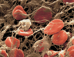
Coagulation (Blood Clotting): A cascade of reactions leads to the formation of a fibrin mesh that traps blood cells, forming a stable clot.
Clotting Cascade (Coagulation)
Tissue Factor (TF): Released by injured tissues.
PF3: Platelet phospholipid that interacts with TF, clotting factors, and calcium ions.
Prothrombin Activator: Converts prothrombin to thrombin.
Thrombin: Converts fibrinogen to insoluble fibrin, forming the clot meshwork.
Equation:
Disorders of Hemostasis
Undesirable Clotting
Thrombus: Clot in an unbroken vessel; can be dangerous if it blocks blood flow.
Embolus: Thrombus that breaks free and travels; can cause blockages elsewhere (e.g., pulmonary embolism, stroke).
Bleeding Disorders
Thrombocytopenia: Low platelet count, leading to spontaneous bleeding and petechiae (small purplish spots).
Hemophilia: Hereditary disorder with missing clotting factors, causing prolonged bleeding.
Blood Groups and Transfusions
Blood Loss and Transfusions
Significant Blood Loss: Loss of 15–30% causes weakness; over 30% can cause shock and be fatal.
Transfusions: Used to treat severe blood loss, anemia, or thrombocytopenia.
Human Blood Groups
Antigens: Genetically determined proteins on RBC membranes; recognized as foreign if not "self".
Antibodies: Proteins that bind foreign antigens, causing agglutination and lysis.
Blood Typing: Mixing blood with anti-A and anti-B sera to detect agglutination.
ABO Blood Group
Type A: Antigen A present.
Type B: Antigen B present.
Type AB: Both antigens present (universal recipient).
Type O: Neither antigen present (universal donor).
Antibodies: IgM type, do not cross placenta.
Rh Blood Group
Rh-positive: Rh antigen present.
Rh-negative: Rh antigen absent; antibodies form only after exposure to Rh-positive blood.
Hemolytic Disease of the Newborn: Occurs when an Rh-negative mother carries an Rh-positive fetus; prevented by RhoGAM injection.
Developmental Aspects of Blood
Fetal and Neonatal Blood
Sites of Formation: Fetal liver and spleen initially; bone marrow takes over by the seventh month.
Fetal Hemoglobin: Differs from adult hemoglobin; higher affinity for oxygen.
Physiologic Jaundice: Occurs in infants when the liver cannot process hemoglobin breakdown products quickly enough.
Blood Disorders Across the Lifespan
Congenital Defects: Hemolytic anemias, hemophilia.
Leukemias: Most common in the very young and very old.
Aging: Increased risk for anemia and clotting disorders in older adults.
