 Back
BackComprehensive Study Notes: Histology (Ch. 4) – Tissue Types and Their Characteristics
Study Guide - Smart Notes
 Tailored notes based on your materials, expanded with key definitions, examples, and context.
Introduction to Tissues & Histology
Overview of Tissue Types
Histology is the scientific study of tissues, focusing on their structure, function, and organization. The human body is composed of four primary tissue types, each with distinct roles and characteristics. Tissues are groups of similar cells working together, often with an associated extracellular matrix (ECM) that provides structural and functional support.
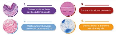
Epithelial Tissue: Covers surfaces, lines cavities, and forms glands.
Connective Tissue: Most abundant and diverse, with a prominent ECM.
Muscle Tissue: Contracts to allow movement.
Nervous Tissue: Detects stimuli and transmits electrical signals.
Histological Techniques
Tissues are typically transparent under a microscope and require staining with dyes to visualize cellular structures. Histologists use various stains to highlight different components, allowing for detailed examination of tissue architecture and pathology.

Lesson Map: Tissue Organization
The study of tissues is organized into four main categories: epithelial, connective, muscle, and nervous tissues. Each category is further subdivided based on structure and function.
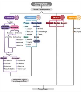
Epithelial Tissue
Introduction and Functions
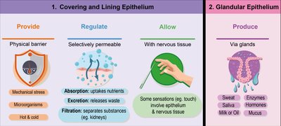
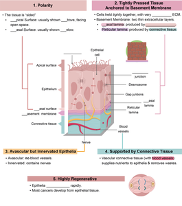
Epithelial tissue forms sheets of tightly packed cells that cover body surfaces, line internal cavities and ducts, and form glands. It is always adjacent to connective tissue, separated by a basement membrane. Epithelial tissues are classified based on their structure and function, including protection, absorption, secretion, and sensation.
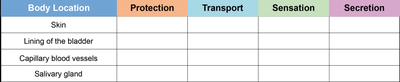
Functional Classification
Body Location | Protection | Transport | Sensation | Secretion |
|---|---|---|---|---|
Skin | X | |||
Lining of the bladder | X | |||
Capillary blood vessels | X | |||
Salivary gland | X |
Characteristics of Epithelial Tissue
Epithelial tissues share several key characteristics:
Polarity: Distinct apical (top) and basal (bottom) surfaces.
Cellularity: Densely packed cells with minimal ECM.
Avascular but Innervated: No blood vessels, but supplied by nerves.
Supported by Connective Tissue: Anchored to a basement membrane.
High Regenerative Capacity: Rapid cell division for repair and renewal.
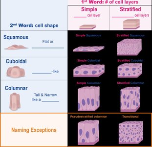
Structural Classification: Layers and Shapes
Epithelial tissues are classified by the number of cell layers and the shape of the cells:
Simple: One cell layer thick.
Stratified: Multiple cell layers.
Squamous: Flat cells.
Cuboidal: Cube-shaped cells.
Columnar: Tall, column-like cells.
Types of Epithelial Tissue
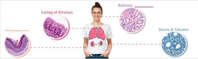
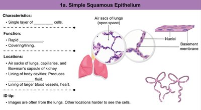
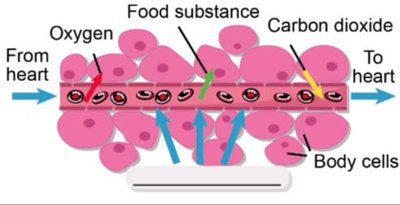
Simple Squamous Epithelium: Single layer of flat cells; allows rapid diffusion and filtration. Found in air sacs of lungs, lining of blood vessels, and serous membranes.
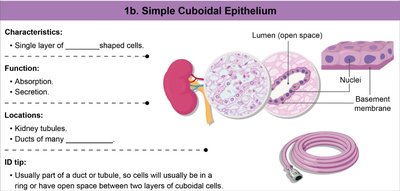
Simple Cuboidal Epithelium: Single layer of cube-shaped cells; functions in absorption and secretion. Located in kidney tubules and ducts of glands.
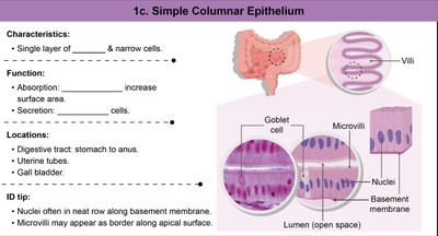
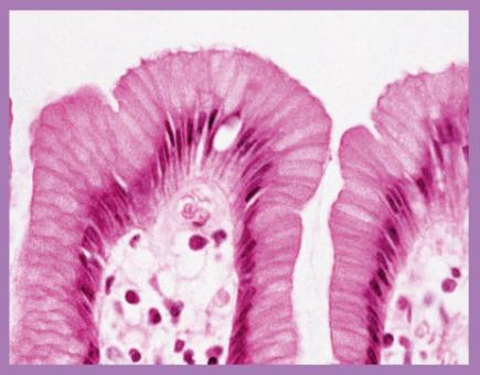
Simple Columnar Epithelium: Single layer of tall, narrow cells; specialized for absorption (with microvilli) and secretion (may contain goblet cells). Found in the digestive tract and gall bladder.
Pseudostratified Columnar Epithelium: Appears layered but all cells touch the basement membrane; often ciliated and contains goblet cells. Located in upper respiratory tract.
Stratified Squamous Epithelium: Multiple layers of flat cells; provides protection against abrasion. Keratinized type forms the skin, non-keratinized lines moist surfaces (mouth, esophagus).
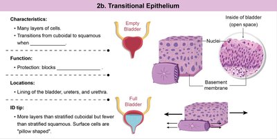
Transitional Epithelium: Multiple layers that change shape; found in the urinary bladder, allowing for stretching.
Glandular Epithelium
Glandular epithelial tissue forms glands that secrete substances. Glands are classified as exocrine (secrete into ducts or onto surfaces) or endocrine (secrete hormones into the bloodstream). Goblet cells are a common unicellular exocrine gland, producing mucus for lubrication and protection.
Connective Tissue
Introduction and General Structure
Connective tissue is the most abundant and diverse tissue class, characterized by a prominent ECM composed of ground substance and protein fibers. All connective tissues originate from embryonic mesenchyme and have cells that occupy less space than the ECM.
Functions of Connective Tissue
Support and structural framework
Protection of organs
Binding and connecting tissues
Storage of energy (as fat)
Transport of substances (blood)
Classification of Connective Tissue
Connective Tissue Proper: Includes loose (areolar, reticular, adipose) and dense (regular, irregular, elastic) types.
Specialized Connective Tissue: Includes cartilage, bone, blood, and lymph.
Connective Tissue Proper: Loose and Dense Types
Loose Connective Tissue: Loosely arranged fibers; supports and cushions organs. Types include areolar, reticular, and adipose tissue.
Dense Connective Tissue: Densely packed fibers; provides strength and resistance to stretching. Types include dense regular, dense irregular, and elastic tissue.
Protein Fibers in Connective Tissue
Collagen Fibers: Strong, flexible, and resist stretching.
Reticular Fibers: Thin, branched, and form supportive networks.
Elastic Fibers: Allow tissues to stretch and recoil.
Muscle Tissue
Types and Functions
Muscle tissue is specialized for contraction and movement. There are three types:
Skeletal Muscle: Voluntary, striated, multinucleated; attaches to bones for body movement.
Cardiac Muscle: Involuntary, striated, branched, with intercalated discs; found in the heart.
Smooth Muscle: Involuntary, non-striated, spindle-shaped; found in walls of hollow organs.
Nervous Tissue
Structure and Function
Nervous tissue is specialized for the generation and transmission of electrical impulses. It is found in the brain, spinal cord, and nerves. Composed of two main cell types:
Neurons: Conduct electrical impulses.
Neuroglia (Glial Cells): Support, protect, and nourish neurons.
Serous Membranes
Organization and Function
Serous membranes (serosa) are thin sheets of tissue that form double-layered membranes around organs in body cavities. They consist of a visceral layer (attached to organs) and a parietal layer (attached to cavity walls), with serous fluid in between to reduce friction during organ movement.
Major Serous Membranes
Pleura: Surrounds the lungs.
Pericardium: Surrounds the heart.
Peritoneum: Surrounds abdominal organs.
Summary Table: Epithelial Tissue Types
Type | Structure | Function | Location |
|---|---|---|---|
Simple Squamous | Single layer, flat cells | Diffusion, filtration | Lungs, blood vessels |
Simple Cuboidal | Single layer, cube-shaped | Absorption, secretion | Kidney tubules, glands |
Simple Columnar | Single layer, tall cells | Absorption, secretion | Digestive tract |
Pseudostratified Columnar | Single layer, appears multilayered | Secretion, movement of mucus | Respiratory tract |
Stratified Squamous | Multiple layers, flat cells | Protection | Skin, mouth, esophagus |
Transitional | Multiple layers, variable shape | Stretching | Urinary bladder |
