Back
BackDevelopment and Heredity: Human Anatomy & Physiology Chapter 27 Study Notes
Study Guide - Smart Notes
 Tailored notes based on your materials, expanded with key definitions, examples, and context.
Development and Heredity
Overview of Human Development
Human development begins at fertilization and continues throughout life, encompassing a series of complex changes in form and function. The field of Developmental Biology studies these changes, while Embryology focuses on the events of the prenatal period (approximately 38 weeks in utero). The Postnatal Period refers to the time from birth through all subsequent life stages.
The Process of Prenatal Development
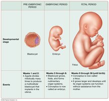
Prenatal development is divided into three main stages:
Pre-Embryonic Period (Weeks 1–2): Begins with fertilization, formation of the zygote, and rapid mitotic divisions to form a blastocyst.
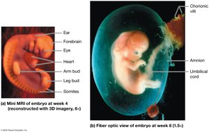
Embryonic Period (Weeks 3–8): The conceptus is now called an embryo; organ systems begin rudimentary formation.
Fetal Period (Weeks 9–38): The embryo is now a fetus; organ systems mature and the fetus grows in size and complexity.
Infertility and Assisted Reproductive Technology (ART)
Infertility is defined as the inability to achieve pregnancy after one year of unprotected intercourse. Causes in males often involve low sperm count, while in females, hormonal imbalances, ovary diseases, or uterine tube/uterus conditions are common. Assisted Reproductive Technology (ART) includes methods such as:
Intrauterine Insemination (IUI): Sperm are placed directly into the uterus.
Gamete Intrafallopian Transfer (GIFT): Sperm and oocytes are placed into the uterine tube.
Zygote Intrafallopian Transfer (ZIFT): Zygote is placed into the uterine tube after fertilization outside the body.
In Vitro Fertilization (IVF): Fertilization occurs outside the body, and the embryo is transferred to the uterus.
The Postnatal Period
The postnatal period is divided into five stages:
Neonatal Period: Birth to 1 month
Infancy: 1 month to 2 years
Childhood: Until puberty
Adolescence: Approximately ages 10–19
Adulthood (Maturity): End of adolescence to death; includes senescence (degeneration of tissues/organs)
Fertilization and Early Embryonic Development
Fertilization
Fertilization marks the beginning of the pre-embryonic period. Meiosis produces haploid gametes (1n, 23 chromosomes). When sperm and oocyte combine, a diploid zygote (2n, 46 chromosomes) forms. Sperm are delivered in semen, which contains substances that facilitate sperm survival and motility in the female reproductive tract.
Only a fraction of the 40–750 million sperm reach the oocyte due to barriers such as acidic vaginal secretions and cervical mucus.
High estrogen levels produce thin cervical mucus (promotes motility); high progesterone produces thick mucus (impedes motility).
Sperm are motile due to their flagellum; oocytes are viable for 24 hours post-ovulation, and sperm can survive up to 5 days in the female tract.
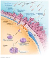
Steps of Fertilization
Capacitation: Sperm undergo changes to become fully motile and able to penetrate the oocyte.
Acrosomal Reaction: Enzymes are released to penetrate the corona radiata and zona pellucida.
Sperm-Oocyte Fusion: Sperm binds to the oocyte membrane, triggering the cortical reaction to prevent polyspermy.
Completion of Meiosis II: The oocyte completes meiosis, forming the female pronucleus.
Amphimixis: Male and female pronuclei fuse, forming the diploid zygote.
Cleavage and Blastocyst Formation
About 30 hours after fertilization, the zygote undergoes cleavage—rapid mitotic divisions producing smaller cells called blastomeres. By day 3, a 16-cell morula forms, which then becomes a blastocyst with an outer trophoblast (forms placenta) and inner cell mass (forms embryo). Monozygotic twins arise from early separation of blastomeres; dizygotic twins result from two separate zygotes.
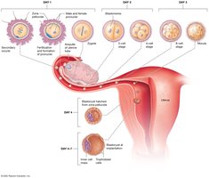
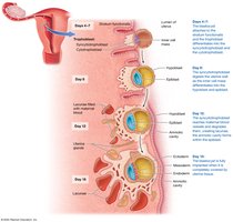
Implantation
Implantation occurs 4–7 days after fertilization as the blastocyst attaches to and invades the endometrium. The trophoblast differentiates into the cytotrophoblast and syncytiotrophoblast (which secretes human chorionic gonadotropin (hCG) to maintain pregnancy). By day 16, the blastocyst is fully embedded in the uterine wall.
Embryonic Development: Germ Layers and Organogenesis
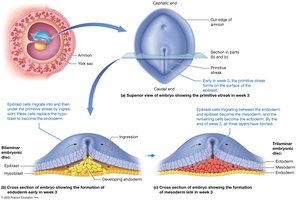
Formation of the Germ Layers (Gastrulation)
During week 3, gastrulation transforms the bilaminar embryonic disc into a trilaminar disc with three primary germ layers:
Ectoderm: Forms the epidermis and nervous system
Mesoderm: Forms muscle, bone, connective tissue, and cardiovascular system
Endoderm: Forms the epithelial lining of the digestive, respiratory, and urinary tracts
Organogenesis
Organogenesis is the process by which the three germ layers differentiate into organs and organ systems. Key events include:
Ectoderm: Forms the neural tube (brain and spinal cord), sense organs, and epidermis.
Mesoderm: Forms the notochord, somites (which become vertebrae, dermis, and skeletal muscle), kidneys, gonads, and cardiovascular system.
Endoderm: Forms the lining of the digestive and respiratory tracts, and several glands.
Placentation and Fetal Development
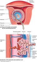
Placentation
Placentation is the formation of the placenta, a temporary organ that facilitates exchange of nutrients, gases, and wastes between mother and fetus. The umbilical cord connects the fetus to the placenta, containing two arteries and one vein surrounded by Wharton's jelly.
Fetal Development
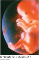
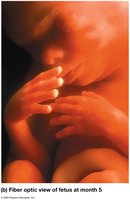
The fetal period is marked by rapid growth and maturation of tissues and organs. Major events by month include:
Month 3: Body lengthens, ossification begins, genitals distinguishable
Month 4: Rapid growth, joints form, heartbeat audible
Month 5: Hair (lanugo) and vernix caseosa develop, mother feels movement (quickening)
Month 6–7: Eyelids open, fat accumulates, lungs produce surfactant, testes descend in males
Month 8–9: Organs mature, lanugo shed, fetus reaches full-term
Maternal Changes During Pregnancy and Parturition
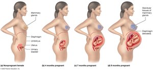
Maternal Changes During Pregnancy
Pregnancy is divided into three trimesters. Hormonal changes include increased hCG, estrogens, and progesterone to maintain the uterine lining. Anatomical and physiological changes affect nearly every system, including increased blood volume, cardiac output, and changes in the position of abdominal organs.
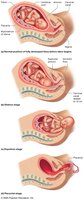
Parturition (Labor and Delivery)
Labor consists of three stages:
Dilation Stage: Cervix dilates to 10 cm; amniotic sac may rupture
Expulsion Stage: Delivery of the newborn; strong uterine contractions and maternal effort
Placental Stage: Delivery of the placenta and membranes (afterbirth)
Neonatal Changes and Heredity
Changes in the Newborn
The neonatal period (first 4 weeks) involves adaptation to life outside the womb. The Apgar score assesses newborn health based on skin color, pulse, respiration, muscle activity, and reflexes. Circulatory and respiratory systems undergo significant changes, and passive immunity is provided by maternal antibodies.
Introduction to Heredity
Genes are DNA segments coding for proteins; alleles are variants of a gene. Heredity is the transmission of genetic traits from parents to offspring. The genome consists of 46 chromosomes (23 pairs), including autosomes and sex chromosomes. Genotype is the genetic makeup; phenotype is the physical expression of traits.
Patterns of Inheritance
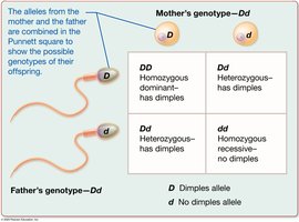
Autosomal Dominant-Recessive: Traits determined by dominant or recessive alleles (e.g., dimples, freckles).
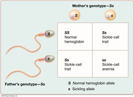
Incomplete Dominance: Heterozygotes show intermediate phenotypes (e.g., sickle-cell trait).
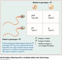
Multiple-Allele Traits: Traits influenced by more than two alleles (e.g., ABO blood group).
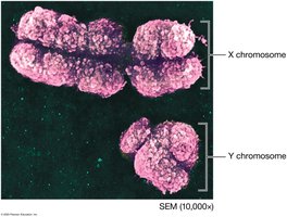
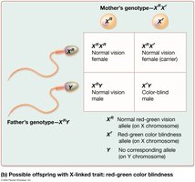
Sex-Linked Traits: Traits carried on X or Y chromosomes (e.g., color blindness, hemophilia).
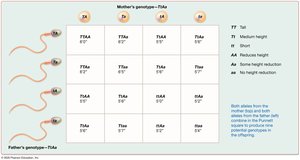
Polygenic Inheritance: Traits influenced by multiple genes (e.g., height, skin color).
Multifactorial Inheritance: Traits influenced by genes and environment.
Genotype | Phenotype |
|---|---|
Blood type A | |
Blood type A | |
Blood type B | |
Blood type B | |
Blood type AB | |
Blood type O |
Prenatal and Newborn Genetic Screening
Genetic screening can detect disorders such as Down syndrome, sickle-cell anemia, and hemophilia. Amniocentesis and chorionic villus sampling are procedures used to obtain fetal cells for genetic analysis, typically guided by ultrasound. Newborn screening involves blood tests to identify metabolic or genetic disorders early.