Back
BackDevelopment and Inheritance: From Fertilization to Genetics
Study Guide - Smart Notes
 Tailored notes based on your materials, expanded with key definitions, examples, and context.
An Introduction to Development and Inheritance
Overview of Development and Inheritance
Development is the gradual modification of anatomical structures and physiological characteristics from fertilization to maturity. Inheritance refers to the transfer of genetic material from one generation to the next, determining the traits of offspring. The study of these processes is essential for understanding human growth, reproduction, and genetic diversity.
Developmental Stages
Embryonic and Fetal Development
Development is divided into several key stages:
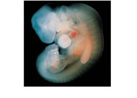
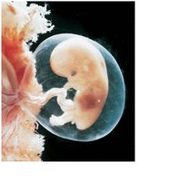
Embryonic Development: Occurs during the first two months after fertilization. The study of these events is called embryology.
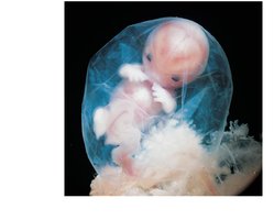
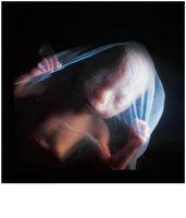
Fetal Development: Begins at the start of the ninth week and continues until birth.
Prenatal Development: Encompasses both embryonic and fetal stages.
Postnatal Development: Begins at birth and continues to maturity.
Fertilization
The Process of Fertilization
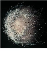
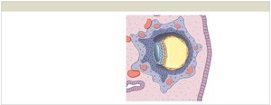
Fertilization is the fusion of two haploid gametes (sperm and oocyte), each containing 23 chromosomes, to form a zygote with 46 chromosomes. This process initiates development and involves several steps:
Spermatozoon: Delivers paternal chromosomes to the fertilization site, traveling a considerable distance and being highly streamlined.
Oocyte: Provides organelles, inclusions, nourishment, and genetic programming for early development.
Capacitation: Sperm must undergo changes in the female reproductive tract to become capable of fertilizing the oocyte.
Acrosomal Enzymes: Hyaluronidase and acrosin help sperm penetrate the protective layers around the oocyte.
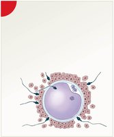
Oocyte Activation: Fusion of sperm and oocyte membranes triggers completion of meiosis II in the oocyte.
Prevention of Polyspermy: The cortical reaction inactivates sperm receptors and hardens the zona pellucida to prevent entry of additional sperm.
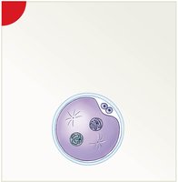
Pronuclei Fusion (Amphimixis): The male and female pronuclei fuse, completing fertilization and forming a zygote.
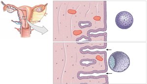
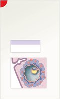
Cleavage: A series of rapid cell divisions producing blastomeres.
Early Embryonic Development
Cleavage and Blastocyst Formation
After fertilization, the zygote undergoes cleavage, a series of mitotic divisions that produce smaller cells called blastomeres. The stages include:
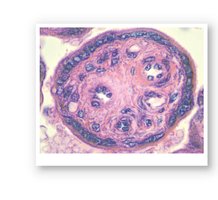
Morula: A solid ball of cells formed after three days of cleavage.
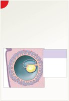
Blastocyst: A hollow ball with an inner cavity (blastocoele), consisting of an outer trophoblast and an inner cell mass.
Trophoblast: Outer layer providing nutrients and later forming part of the placenta.
Inner Cell Mass: Cluster of cells that will form the embryo.
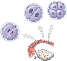
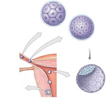
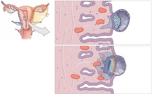
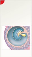
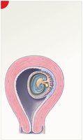
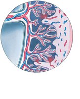
Implantation
Implantation occurs about seven days after fertilization, when the blastocyst adheres to the uterine lining. The trophoblast differentiates into cellular and syncytial layers, with the latter secreting enzymes to facilitate implantation.
Gastrulation and Germ Layer Formation
Formation of Germ Layers
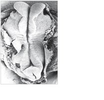
Gastrulation is the process by which the inner cell mass forms three primary germ layers:
Ectoderm: Forms the epidermis, nervous system, and some endocrine structures.
Mesoderm: Forms muscle, bone, connective tissue, cardiovascular system, and more.
Endoderm: Forms the lining of the digestive and respiratory tracts, and associated organs.
Extraembryonic Membranes and Placenta
Formation and Function
Four extraembryonic membranes support embryonic and fetal development:
Yolk Sac: Early site of blood cell formation.
Amnion: Encloses the embryo in amniotic fluid for protection.
Allantois: Contributes to the formation of the urinary bladder.
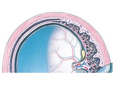
Chorion: Forms the placenta, facilitating nutrient and gas exchange.
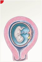
Placental Structure and Circulation
The placenta is a complex organ that supports fetal development by exchanging nutrients, gases, and wastes between maternal and fetal blood. It also produces hormones essential for pregnancy maintenance.
Umbilical Cord: Connects fetus to placenta, containing blood vessels for nutrient and waste exchange.
Placental Hormones: Includes hCG, hPL, placental prolactin, relaxin, progesterone, and estrogens.
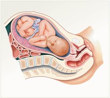
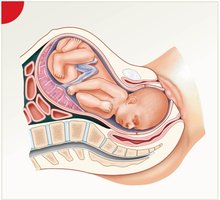
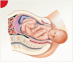
Gestation: Trimesters and Major Events
First Trimester
The first trimester includes cleavage, implantation, placentation, and embryogenesis. It is the most critical period, with the highest risk of developmental abnormalities.
Second and Third Trimesters
During the second trimester, organs and systems continue to develop, and the fetus grows rapidly. The third trimester is characterized by further growth, maturation of organ systems, and preparation for birth.
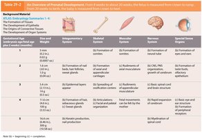
Overview Table of Prenatal Development
The following table summarizes the major events in prenatal development across different organ systems:
Gestational Age | Integumentary System | Skeletal System | Muscular System | Nervous System | Special Senses |
|---|---|---|---|---|---|
1-4 weeks | Formation of skin layers | Formation of somites | Formation of myotomes | CNS, PNS formation | Formation of eyes and ears |
5-8 weeks | Development of appendages | Ossification begins | Muscle mass increases | Brain regions develop | Eye and ear structures mature |
9-12 weeks | Keratin production | Bone growth | Muscle differentiation | Reflexes develop | Functional eyes and ears |
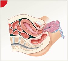
Labor and Delivery
Stages of Labor
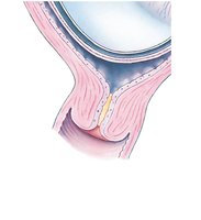
Labor is the process of parturition, or childbirth, and consists of three stages:
Dilation Stage: Cervix dilates, amniochorionic membrane ruptures ("water breaks").
Expulsion Stage: Fetus is delivered through the birth canal.
Placental Stage: Placenta is expelled from the uterus.
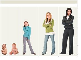
Postnatal Life Stages
Stages and Major Events
Postnatal development is divided into several stages:
Neonatal Period: Birth to 1 month; organ systems begin independent function.
Infancy: 1 month to 2 years; rapid growth and development.
Childhood: 2 years to adolescence; continued growth and maturation.
Adolescence: Puberty to maturity; sexual and physical maturation.
Maturity (Senescence): Aging and decline in functional capacity.
Genetics and Inheritance
Basic Principles
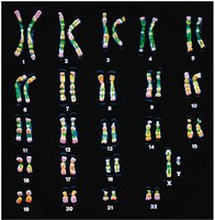
Inheritance is governed by the transmission of genes on chromosomes. Each somatic cell contains 23 pairs of chromosomes (22 autosomes and 1 pair of sex chromosomes). The genotype is the genetic makeup, while the phenotype is the physical expression of those genes.
Patterns of Inheritance
Simple Inheritance: Phenotype determined by a single pair of alleles.
Polygenic Inheritance: Multiple genes interact to determine phenotype.
Sex-Linked Inheritance: Traits determined by genes on sex chromosomes, especially the X chromosome.
Dominance Relationships: Includes strict dominance, incomplete dominance, and codominance.
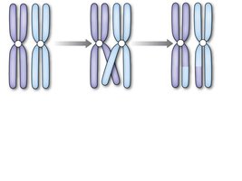
Genetic Variation and Recombination
Genetic diversity arises from random assortment of chromosomes during meiosis and from crossing over, where homologous chromosomes exchange segments. Mutations and chromosomal abnormalities can also contribute to variation and disease.
Human Genome and Genetic Disorders
The Human Genome Project mapped thousands of human genes, aiding in the understanding of genetic diseases and inheritance patterns. Karyotyping is used to detect chromosomal abnormalities.