 Back
BackDevelopment and Inheritance: Human Embryo and Fetal Development
Study Guide - Smart Notes
 Tailored notes based on your materials, expanded with key definitions, examples, and context.
Gestation and Stages of Human Development
Overview of Gestation
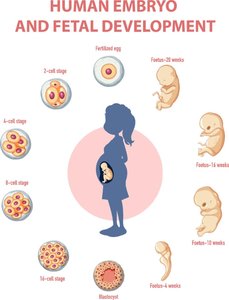
Gestation is the period during which the embryo and fetus develop in the uterus, culminating in birth. It is divided into three main stages:
Pre-embryonic stage: Fertilization to week 2
Embryonic stage: Weeks 3-8
Fetal stage: Week 9 to birth
Fertilization
Mechanism of Fertilization
Fertilization is the fusion of the sperm and oocyte pronuclei, resulting in a diploid embryo.
Fusion: nsperm + noocyte = 2nembryo
Vaginal environment: The acidic pH (3.8) kills millions of sperm immediately; cervical mucus blocks more.
Survival: Sperm live 3-5 days; oocyte survives ~24 hours.
Capacitation of Sperm
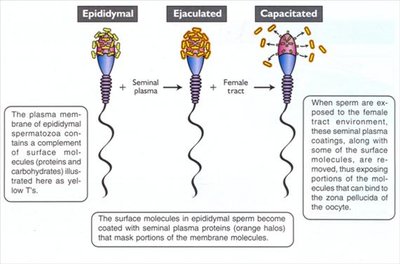
Capacitation is the process by which sperm gain the ability to fertilize the oocyte.
Priming: The female reproductive tract modifies sperm, improving motility and thinning the acrosomal membrane.
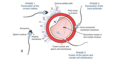
Acrosomal reaction: Acrosomal enzymes interact with the zona pellucida, allowing sperm penetration.
Oocyte Layers and Sperm Penetration
The oocyte is surrounded by two layers:
Zona pellucida: Inner glycoprotein membrane, reacts with lysosomes.
Corona radiata: Outer layer of granulosa cells, digested by multiple acrosomes.
Chemotaxis: Oocyte releases chemoattractants to guide sperm.
Prevention of Polyspermy
Fast-Block Mechanism
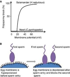
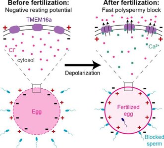
To prevent polyspermy (fertilization by more than one sperm), the oocyte employs a fast-block mechanism:
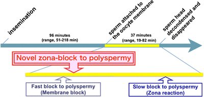
Na+ permeability: Depolarizes cell membrane for ~1 minute, blocking additional sperm entry.
Slow-Block Mechanism (Cortical Reaction)
The slow-block involves the cortical reaction:
Zonal inhibiting protein: Causes sperm to release and destroys sperm receptors on oocyte.
Mucopolysaccharides: Join with zona pellucida to form an impenetrable fertilization membrane.
Zygote Formation and Early Cleavage
Zygote Formation
After fertilization:
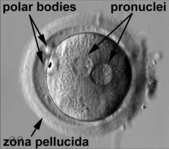
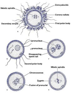
Meiosis II: Metaphase II begins, polar body ejected, meiosis completed.
Pronuclei: Form and chromosomes intermingle, creating a diploid zygote.
Mitosis: Begins, leading to cellular division.
Twins: Fraternal twins result from fertilization of two separate oocytes.
Pre-Implantation Development
Cleavage and Blastocyst Formation
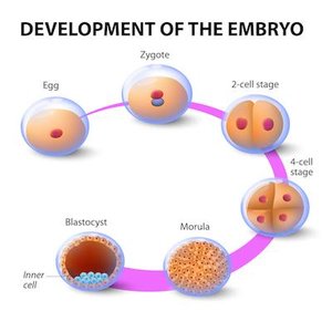
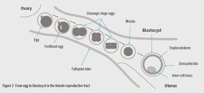
Early embryonic development involves several stages:
Blastomere: Initial cellular division.
Morula: 16-cell stage, enters uterus.
Blastocyst: Blastocoel visible, cellular differentiation begins.
Blastocyst Structure
Blastocoel: Fluid-filled cavity.
Inner cell mass: Embryo, totipotent cells.
Trophoblasts: Outer layer, nourish egg, form chorionic sac and fetal placenta.
Hatching: Embryo sheds zona pellucida before implantation.
Implantation
Mechanism of Implantation
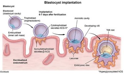
Trophoblasts: Facilitate implantation; 50-70% fail to implant.
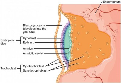
Syncytiotrophoblast: Fused trophoblasts digest endometrial cells and secure blastocyst.
Hormonal Changes and Pregnancy Detection
Uterine mucosa: Envelops blastocyst after syncytiotrophoblast fusion.
hCG secretion: Trophoblasts secrete human chorionic gonadotropin (hCG).
Corpus luteum: Produces progesterone and estrogen.
Pregnancy test: Detects hCG a few days after implantation.
Problematic Implantations
Ectopic Pregnancy and Placenta Previa
Ectopic pregnancy: Implantation outside uterus (tubal, ovarian, abdominal).
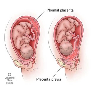
Placenta previa: Embryo implants in inferior uterus, placenta covers internal os (cervical region).
Symptoms: Profuse bleeding at 24 weeks, antepartum hemorrhage.
Embryonic Membranes
Formation and Function of Membranes
Embryonic membranes are essential for protection and nourishment:
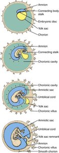
Amnion: Develops from trophoblasts, surrounds amniotic fluid and inner cell mass.
Yolk sac: Hypoblast layer, may develop into germ or blood cells.
Allantois: Joins yolk sac, becomes part of urinary bladder and umbilical cord.
Chorion: Fusion of yolk sac and allantois, surrounds all other membranes.
Chorion and Placenta Formation
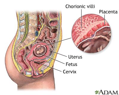
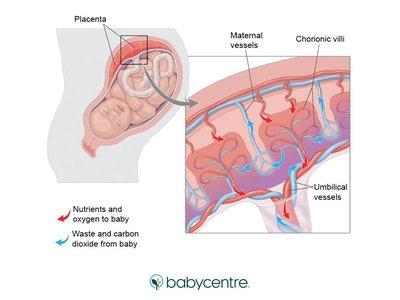
Chorionic villi: Burrow into endometrium, join fetal mesenchymal cells, develop placenta.
Embryogenesis and Germ Layers
Gastrulation and Germ Layer Formation
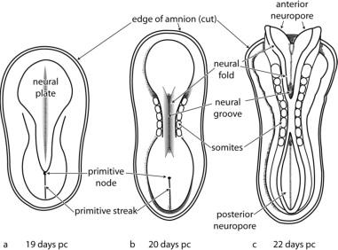
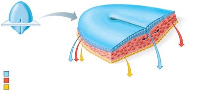
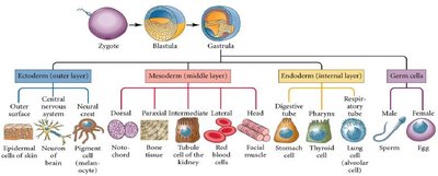
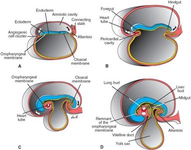
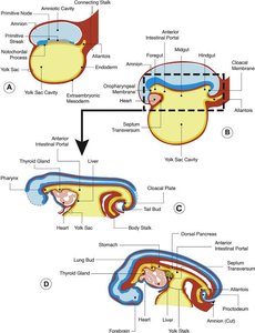
Embryogenesis begins at week 3, with the embryonic disc forming three germ layers via gastrulation:
Primitive streak: Emits growth factors, leads to germ layer formation.
Germ layers: Ectoderm (nervous tissue, skin), Mesoderm (muscle, connective tissue), Endoderm (digestive system lining).
Development of Placenta
Placental Structure and Function
Chorion: Initiates placental development around 14-16 weeks.
Umbilical arteries: Carry blood away from fetus' heart (low O2).
Umbilical veins: Carry blood toward fetus' heart (high O2).
Blood separation: Prevents maternal cytotoxic T cell attack on fetus.
Embryonic Milestones and Organogenesis
Key Developmental Events
4th week: Heart tubes form.
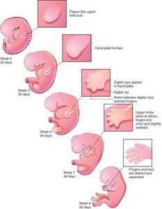
4-5 weeks: Eye pit, limb buds, rudimentary pulmonary system.
6th week: Uncontrolled limb movements, intestines develop, fingers created by apoptosis.
7th week: Facial features begin.
8th week: Organogenesis, embryo is 3 cm and 8 g.
Organogenesis and Brain Development
Neurulation and Brain Vesicle Formation
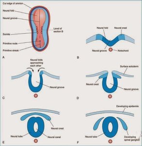
Neurulation: Neural plate forms neural fold, then neural tube, which merges with notochord.
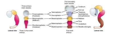
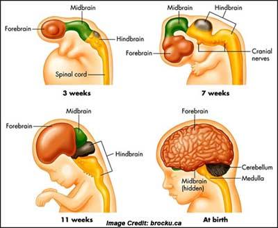
Brain vesicles: Develop over 4-5 weeks; folate is crucial for preventing neural tube defects (e.g., spina bifida, anencephaly).
Three major vesicles: Forebrain (prosencephalon), midbrain (mesencephalon), hindbrain (rhombencephalon).
Fetal Development
Fetal Stage and Organ System Maturation
The fetal stage extends from the 9th week until birth, with newborns capable of life outside the womb.
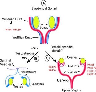
9th-12th week: Sexual differentiation, bipotential gonads influence Müllerian or Wolffian duct disintegration.
Blood shunts: Ductus venosus (bypasses liver), foramen ovale (bypasses lungs), ductus arteriosus (develops pulmonary circuit).
Waste removal: All fetal wastes diffuse through placenta into maternal circulation.
Fetal Milestones
9-12 weeks: Frequent fetal movement, erythropoiesis by fetal bone marrow, amniotic breathing, urine production, eyes developed but shut.
13-16 weeks: Special sense organs, kidneys well-formed, intestines accumulate meconium (amniotic fluid, cellular debris, mucus, bile).
16-20 weeks: Fetal movements, fetal positioning.
21-30 weeks: Myelination, surfactant production, testes descend.
After 30 weeks: Subcutaneous fat builds up.
Example: Meconium is the first stool of the fetus, composed of ingested amniotic fluid, cellular debris, mucus, and bile. Additional info: Fetal development is marked by progressive organ system maturation and increasing viability outside the womb.
