 Back
BackDigestive System and Blood: Anatomy & Physiology Study Guide
Study Guide - Smart Notes
 Tailored notes based on your materials, expanded with key definitions, examples, and context.
Digestive System
Overview of Digestive System Anatomy
The digestive system is responsible for breaking down food, absorbing nutrients, and eliminating waste. It consists of a series of organs and accessory structures that work together to accomplish these functions. - Oral Cavity: Includes teeth, tongue, palate, and salivary glands. - Pharynx and Esophagus: Pathways for food to move from the mouth to the stomach. - Stomach: Churns food and begins protein digestion. - Small Intestine: Main site for nutrient absorption. - Large Intestine: Absorbs water and forms feces. 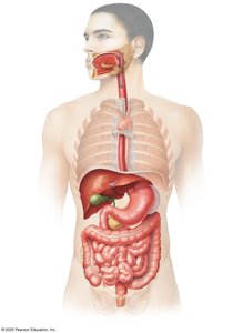
Accessory Digestive Organs
Accessory organs play crucial roles in digestion by producing enzymes, hormones, and other substances. - Salivary Glands: Produce saliva containing lysozyme (antibacterial) and amylase (starch digestion). - Liver: Produces bile, which contains bilirubin (a breakdown product of heme) and aids in fat digestion. - Pancreas: Produces digestive enzymes, bicarbonate (neutralizes acid), and hormones (insulin, glucagon). - Gallbladder: Stores and concentrates bile. 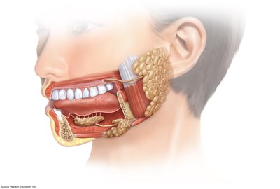
Oral Cavity and Pharynx
The oral cavity is the entry point for food and is involved in mechanical and chemical digestion. - Teeth: Cut, tear, and crush food (mastication). - Tongue: Mixes food with saliva and aids in swallowing (deglutition). - Palate and Uvula: Separate oral and nasal cavities; uvula helps prevent food from entering the nasal passage. - Pharynx: Passageway for food and air. 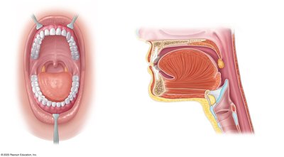
Tooth Anatomy
Teeth are specialized structures for mechanical digestion. - Enamel: Hard, mineralized covering of the crown. - Dentin: Softer mineralized tissue forming most of the tooth. - Cementum: Covers the root, anchoring it to the jawbone. - Pulp Cavity: Contains nerves and blood vessels. - Periodontal Ligaments: Anchor the tooth in the alveolar socket. 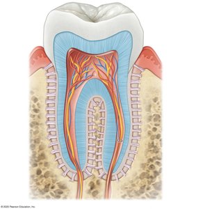
Abdominal Cavity and Mesenteries
The abdominal cavity houses the digestive organs and is supported by mesenteries, which anchor and supply blood to the intestines. - Greater Omentum: Fatty apron covering intestines. - Lesser Omentum: Connects stomach and liver. - Mesenteries: Support and supply intestines. 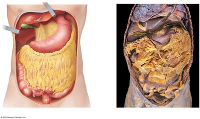
Stomach Anatomy
The stomach is a muscular organ that mixes food with gastric juices. - Regions: Cardia (entry), Fundus (above entry), Body (main part), Pyloric (exit). - Muscle Layers: Longitudinal, circular, and oblique for churning. - Rugae: Folds allowing expansion. - Pyloric Sphincter: Controls passage to the small intestine. 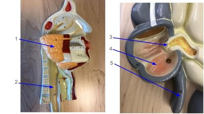
Small Intestine Anatomy
The small intestine is the main site for digestion and absorption. - Duodenum: Receives bile and pancreatic enzymes. - Jejunum: Main site for nutrient absorption. - Ileum: Absorbs remaining nutrients; ends at ileocecal valve. 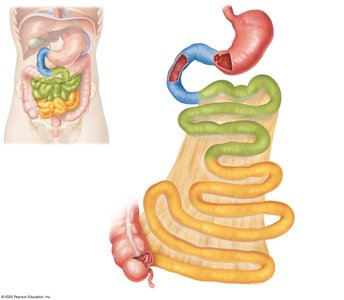
Large Intestine Anatomy
The large intestine absorbs water and forms feces. - Regions: Cecum, ascending colon, transverse colon, descending colon, sigmoid colon, rectum. - Ileocecal Valve: Controls entry from small intestine. - Appendix: Lymphatic tissue. 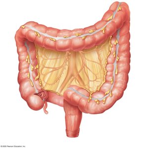
Liver, Gallbladder, and Pancreas Anatomy
These organs are essential for digestion and metabolism. - Liver: Produces bile, processes nutrients. - Gallbladder: Stores bile. - Pancreas: Produces digestive enzymes and hormones. - Bile and Pancreatic Ducts: Deliver secretions to duodenum. 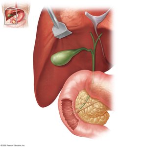
Selected Digestive Hormones
Hormones regulate digestive processes.
Organ | Hormones Produced |
|---|---|
Pancreas | Insulin, Glucagon, Cholecystokinin, GLP-1 |
Stomach | Gastrin, Ghrelin |
Intestine | Various regulatory hormones |
Adipose | Leptin |
Metabolism
Aerobic and Anaerobic Respiration
Cellular respiration is the process of making ATP from nutrients. - Aerobic Respiration: Uses oxygen; occurs in mitochondria; produces ATP, water, and carbon dioxide. - Anaerobic Respiration: Occurs without oxygen; produces ATP and lactic acid (fermentation); lowers blood pH. 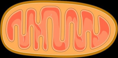
Blood
Components of Blood
Blood is a specialized connective tissue with several components. - Plasma: 50-64% of blood; contains water and plasma proteins. - Buffy Coat: Less than 1%; contains white blood cells (WBCs) and platelets. - Hematocrit: 36-50%; contains red blood cells (RBCs). 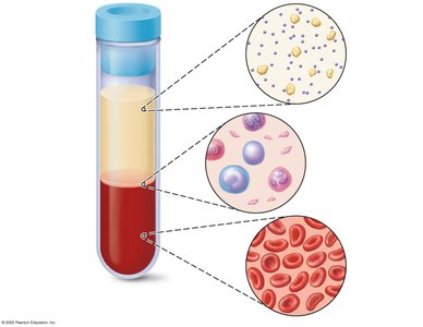
Erythrocyte Structure
Erythrocytes (RBCs) are specialized for oxygen transport. - Hemoglobin: Protein with iron-containing heme groups; binds oxygen. - Structure: Biconcave shape increases surface area for gas exchange. 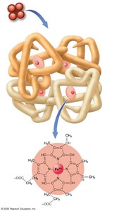
Hematopoiesis
Hematopoiesis is the process of blood cell formation from stem cells. - Hematopoietic Stem Cell: Gives rise to all blood cells. - Myeloid Lineage: Produces erythrocytes, platelets, neutrophils, eosinophils, basophils, monocytes. - Lymphoid Lineage: Produces T and B lymphocytes, natural killer cells. 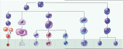
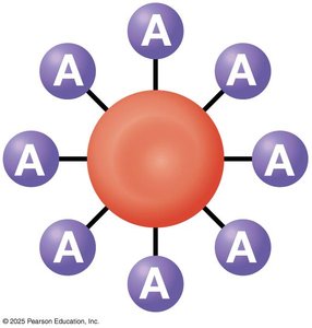
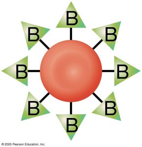
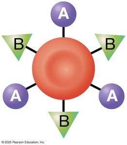
Blood Types
Blood types are determined by antigens on the surface of RBCs.
Blood Type | Antigens | Antibodies | Transfusion Compatibility |
|---|---|---|---|
A | A | Anti-B | Can receive A, O |
B | B | Anti-A | Can receive B, O |
AB | A, B | None | Universal recipient |
O | None | Anti-A, Anti-B | Universal donor |
Leukemia
Leukemia is a cancer of blood-forming tissues. - Lymphocytic Leukemia: Cancerous lymphoid stem cells. - Myeloblastic Leukemia: Cancerous myeloid stem cells. - Acute: Rapid onset and progression. - Chronic: Slower onset and progression.
Additional info:
- The notes cover topics from Ch. 22 (Digestive System), Ch. 23 (Metabolism and Nutrition), Ch. 19 (Blood), and related anatomy and physiology chapters. - Images included are directly relevant to the anatomical and physiological explanations provided.
