 Back
BackDigestive System: Pharynx, Esophagus, Stomach, Liver, Gallbladder, and Pancreas
Study Guide - Smart Notes
 Tailored notes based on your materials, expanded with key definitions, examples, and context.
The Digestive System: Pharynx and Esophagus
The Pharynx
The pharynx serves as a passageway for food, fluids, and air, connecting the mouth to the esophagus and larynx. It is lined with stratified squamous epithelium and contains mucus-producing glands for lubrication and protection.
Muscle Layers: The external muscle consists of two skeletal muscle layers: an inner longitudinal layer and outer pharyngeal constrictors that encircle the wall.
Function: Allows the passage of food, fluids, and air.
The Esophagus
The esophagus is a flat, muscular tube that connects the laryngopharynx to the stomach. It is normally collapsed except during food propulsion.
Location: Pierces the diaphragm at the esophageal hiatus and joins the stomach at the cardial orifice.
Gastroesophageal (cardiac) sphincter: Surrounds the cardial orifice, keeping it closed when food is not being swallowed. Mucus cells protect the esophagus from acid reflux.
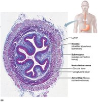
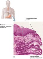
Histology: The esophagus has all four alimentary canal layers. The mucosa contains stratified squamous epithelium, which transitions to simple columnar at the stomach. The muscularis externa transitions from skeletal muscle (superiorly) to smooth muscle (inferiorly). The outermost layer is adventitia (fibrous connective tissue), not serosa.
Clinical Note: Heartburn and GERD
Heartburn: Caused by stomach acid regurgitating into the esophagus, often the first symptom of gastroesophageal reflux disease (GERD).
Causes: Overeating, obesity, pregnancy, running, or hiatal hernia (stomach protrudes above diaphragm).
Complications: Can lead to esophagitis, ulcers, or esophageal cancer.
Digestive Processes of the Mouth, Pharynx, and Esophagus
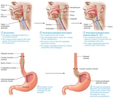
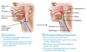
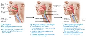
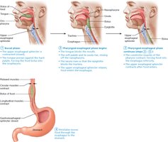
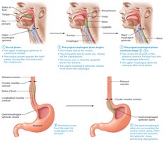
The main function of the pharynx and esophagus is propulsion—moving food from the mouth to the stomach. This process is called deglutition (swallowing), which involves 22 muscle groups and two phases:
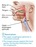
Buccal phase: Voluntary contraction of the tongue.
Pharyngeal-esophageal phase: Involuntary, controlled by the swallowing center in the medulla and lower pons, primarily involving the vagus nerve.
The Stomach
Gross Anatomy of the Stomach
The stomach is a muscular, expandable organ that serves as a temporary storage tank and initiates protein digestion. It converts food into a semi-liquid mixture called chyme.
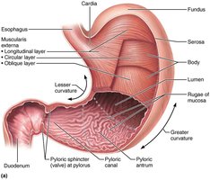
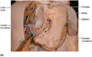
Regions: Cardia, fundus, body, and pyloric part (antrum, canal, pylorus).
Curvatures: Greater curvature (lateral), lesser curvature (medial).
Mesenteries: Lesser omentum (to liver), greater omentum (drapes over intestines).
Volume: Empty stomach holds ~50 ml; can expand to 4 L.
Rugae: Folds in the mucosa when the stomach is empty.
Blood supply: Celiac trunk (gastric and splenic branches); veins drain into hepatic portal system.
Innervation: Sympathetic (thoracic splanchnic nerves via celiac plexus), parasympathetic (vagus nerve).
Microscopic Anatomy of the Stomach
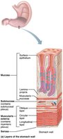
The stomach wall contains the four basic layers, but the muscularis and mucosa are specialized for its digestive functions.
Muscularis externa: Three layers—longitudinal, circular, and oblique (unique to stomach)—allow churning and mixing of chyme.
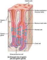
Mucosa: Simple columnar epithelium with mucous cells, producing a two-layer coat of alkaline mucus. Contains gastric pits leading to gastric glands.
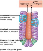
Gastric glands: Secrete gastric juice and contain several cell types:
Mucous neck cells: Secrete thin, acidic mucus.
Parietal cells: Secrete HCl (pH 1.5–3.5) and intrinsic factor (for vitamin B12 absorption).
Chief cells: Secrete pepsinogen (inactive, converted to pepsin by HCl) and lipases.
Enteroendocrine cells: Secrete hormones (gastrin, somatostatin) and paracrines (serotonin, histamine).
Mucosal Barrier and Clinical Imbalances
Mucosal barrier: Protects the stomach from harsh acidic conditions via a thick mucus layer, tight junctions, and rapid cell turnover (every 3–6 days).
Gastritis: Inflammation from breach of the mucosal barrier.
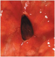
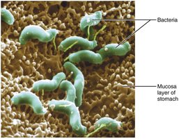
Peptic/gastric ulcers: Erosions in the stomach wall, often caused by Helicobacter pylori or NSAIDs (e.g., aspirin). Severe cases can lead to peritonitis or hemorrhage.
Digestive Processes in the Stomach
Functions: Mechanical and chemical breakdown of food, holding area, delivery of chyme to small intestine, protein denaturation (HCl), enzymatic digestion (pepsin), and absorption of some substances (alcohol, aspirin).
Intrinsic factor: Essential for vitamin B12 absorption; deficiency leads to pernicious anemia.
Regulation of Gastric Secretion
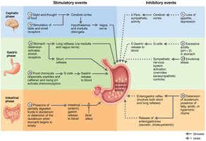
Gastric secretion (>3 L/day) is regulated by neural (vagus nerve increases, sympathetic decreases) and hormonal (gastrin stimulates HCl) mechanisms. Secretion occurs in three phases:
Cephalic phase: Triggered by sight, smell, taste, or thought of food.
Gastric phase: Lasts 3–4 hours; initiated by stomach distension and chemical stimuli (proteins, caffeine, low acidity) that stimulate gastrin release.
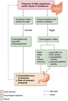
Intestinal phase: Brief stimulation as chyme enters duodenum, followed by inhibition (enterogastric reflex, enterogastrones such as secretin and CCK).
Mechanism of HCl Formation
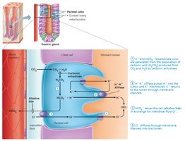
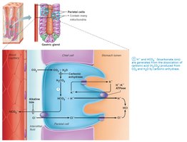
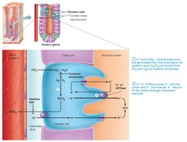
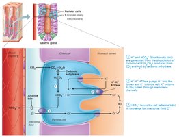
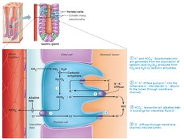
Parietal cells secrete HCl into the stomach lumen via the H+/K+ ATPase (proton pump). Bicarbonate (HCO3−) is exported to the blood, causing an "alkaline tide." Chloride ions join H+ in the lumen to form HCl.
Equation:
Regulation of Gastric Motility and Emptying
Filling response: Stomach stretches to accommodate food via receptive relaxation and gastric accommodation.
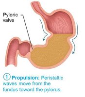
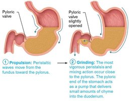
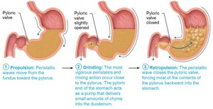
Contractile activity: Peristaltic waves (3/min) move chyme toward the pylorus. Only small particles and liquids pass through the pyloric valve.
Regulation: Duodenum controls gastric emptying via stretch and chemical receptors, enterogastric reflex, and enterogastrones. Fatty chyme slows emptying; carbohydrate-rich chyme moves quickly.
Clinical Note: Vomiting (Emesis)
Causes: Extreme stretching, irritants (toxins, alcohol, spicy food, drugs).
Mechanism: Sensory impulses stimulate the emetic center in the medulla.
Complications: Excessive vomiting can cause dehydration and alkalosis (loss of acid).
Liver, Gallbladder, and Pancreas
The Liver
The liver is the largest gland in the body and has a primary digestive function of producing bile, which emulsifies fats.
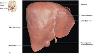
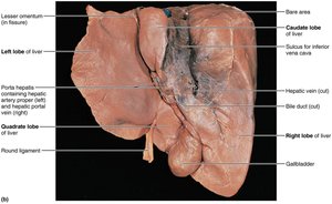
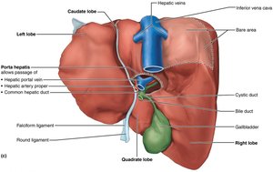
Lobes: Right, left, caudate, quadrate.
Ligaments: Falciform (separates right and left lobes), round ligament (remnant of fetal umbilical vein).
Bile ducts: Common hepatic duct (from liver), cystic duct (to gallbladder), bile duct (union of both).
Microscopic Anatomy of the Liver
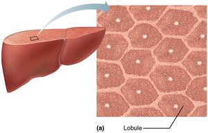
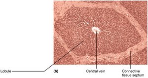
Liver lobules: Hexagonal units made of hepatocyte plates surrounding a central vein. Portal triads at each corner contain a branch of the hepatic artery, portal vein, and bile duct.
Liver sinusoids: Leaky capillaries between hepatocyte plates; blood flows from portal triads to central vein.
Stellate macrophages: Remove debris and old RBCs.
Hepatocyte functions: Produce bile, process nutrients, store vitamins, detoxify blood.
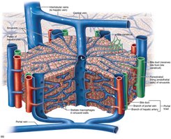
Bile: Composition and Enterohepatic Circulation
Components: Bile salts (fat emulsification), bilirubin (heme breakdown pigment), cholesterol, triglycerides, phospholipids, electrolytes.
Enterohepatic circulation: Bile salts are reabsorbed in the ileum, returned to the liver, and resecreted. About 95% are recycled.
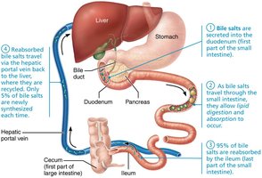
Clinical Note: Liver Disorders
Hepatitis: Inflammation, usually viral, but can be due to toxins or drugs.
Cirrhosis: Chronic inflammation leading to fibrosis and portal hypertension. The liver can regenerate if a portion is removed.
The Gallbladder
The gallbladder stores and concentrates bile, releasing it via the cystic duct into the bile duct during digestion.
Gallstones (biliary calculi): Formed from excess cholesterol or insufficient bile salts, can block bile flow and cause pain or jaundice.
Treatment: Drugs, ultrasound, laser, or surgery.
The Pancreas
The pancreas is both an exocrine and endocrine gland. Its exocrine function is to produce pancreatic juice, which contains digestive enzymes and bicarbonate to neutralize stomach acid.
Acini: Clusters of secretory cells producing enzyme-rich juice.
Duct cells: Secrete bicarbonate-rich fluid.
Islets of Langerhans: Endocrine cells producing insulin and glucagon.
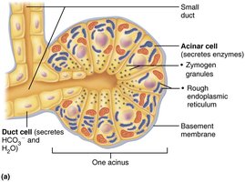
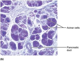
Pancreatic Juice Composition and Enzyme Activation
Contents: 1200–1500 ml/day, alkaline (pH 8), contains electrolytes and digestive enzymes (proteases, amylase, lipases, nucleases).
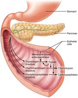
Protease activation: Proteases are secreted in inactive forms and activated in the duodenum by enteropeptidase (e.g., trypsinogen to trypsin).
Bile and Pancreatic Secretion into the Small Intestine
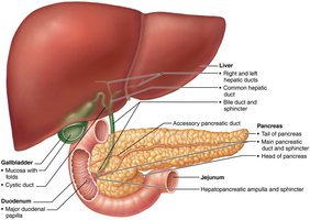
Bile duct and pancreatic duct: Unite at the hepatopancreatic ampulla, opening into the duodenum via the major duodenal papilla. The hepatopancreatic sphincter controls entry.
Accessory pancreatic duct: Empties directly into the duodenum.
Regulation of Bile and Pancreatic Secretion
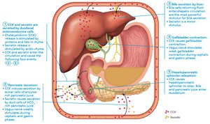
Neural and hormonal controls: Cholecystokinin (CCK) and secretin stimulate secretion in response to chyme entering the duodenum.
Bile secretion: Increased by bile salts returning via enterohepatic circulation and by secretin.
Gallbladder contraction: Stimulated by CCK.
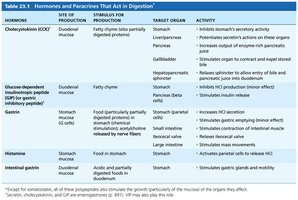
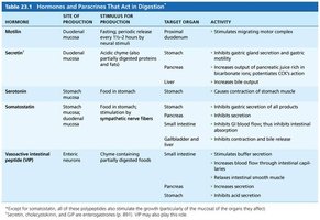
Hormone | Site of Production | Stimulus | Target Organ | Activity |
|---|---|---|---|---|
Cholecystokinin (CCK) | Duodenal mucosa | Fatty chyme | Stomach, liver/pancreas, gallbladder | Inhibits gastric activity, increases pancreatic juice, stimulates bile release |
Secretin | Duodenal mucosa | Acidic chyme | Stomach, pancreas, liver | Inhibits gastric secretion, increases bicarbonate-rich juice, increases bile output |
Gastrin | Stomach mucosa | Food in stomach | Stomach, small intestine, ileocecal valve, large intestine | Increases HCl secretion, stimulates contraction, relaxes ileocecal valve, mass movements |
Somatostatin | Stomach, duodenum | Food in stomach, sympathetic activity | Stomach, pancreas, small intestine, gallbladder/liver | Inhibits gastric secretion, inhibits pancreatic secretion, inhibits GI blood flow, inhibits contraction and bile release |
Additional info: Table summarizes the main hormones and paracrines involved in digestive regulation, their sites of production, stimuli, target organs, and activities.
