 Back
BackDigestive System: Structure, Function, and Disorders
Study Guide - Smart Notes
 Tailored notes based on your materials, expanded with key definitions, examples, and context.
Digestive System Overview
Main and Accessory Organs
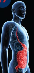
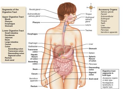
The digestive system consists of a series of organs responsible for the breakdown, absorption, and metabolism of nutrients. It is divided into main organs forming the alimentary canal and accessory organs that aid digestion.
Main organs: Mouth, pharynx, esophagus, stomach, small intestine, large intestine, rectum, and anal canal
Accessory organs: Teeth and tongue, salivary glands (parotid, submandibular, sublingual), liver, gallbladder, pancreas, and vermiform appendix
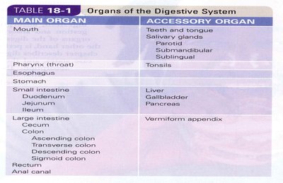
Main Organ | Accessory Organ |
|---|---|
Mouth | Teeth and tongue, Salivary glands (Parotid, Submandibular, Sublingual) |
Pharynx (throat) | Tonsils |
Esophagus | |
Stomach | |
Small intestine (Duodenum, Jejunum, Ileum) | Liver, Gallbladder, Pancreas |
Large intestine (Cecum, Colon, Rectum, Anal canal) | Vermiform appendix |
Primary Mechanisms of the Digestive System
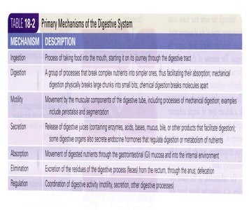
The digestive system operates through several coordinated mechanisms:
Ingestion: Intake of food into the mouth
Digestion: Breakdown of complex nutrients into simpler ones, both mechanically and chemically
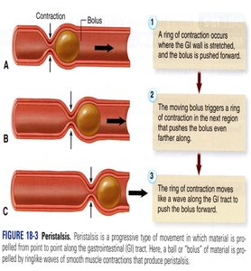
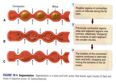
Motility: Movement of food through the digestive tract via muscular contractions (peristalsis and segmentation)
Secretion: Release of digestive juices and enzymes
Absorption: Movement of nutrients from the GI tract into blood or lymph
Elimination: Removal of undigested waste
Regulation: Coordination of digestive activity
Mechanism | Description |
|---|---|
Ingestion | Process of taking food into the mouth |
Digestion | Breakdown of nutrients into simpler forms |
Motility | Movement by muscular components (peristalsis, segmentation) |
Secretion | Release of digestive juices and hormones |
Absorption | Transfer of nutrients into blood/lymph |
Elimination | Excretion of waste |
Regulation | Coordination of digestive processes |
Wall of the Digestive Tract
Four Layers
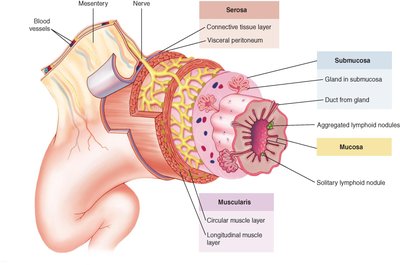
The digestive tract wall is composed of four distinct layers, each with specialized functions:
Mucosa: Innermost layer; contains glands and lymphoid tissue
Submucosa: Connective tissue with blood vessels, nerves, and glands
Muscularis: Smooth muscle layers responsible for motility
Serosa: Outermost layer; connective tissue and visceral peritoneum
Motility: Peristalsis and Segmentation
Peristalsis: Progressive wave-like contractions that propel food through the GI tract
Segmentation: Back-and-forth contractions that mix and break down food
Mouth and Oral Cavity
Structure and Function
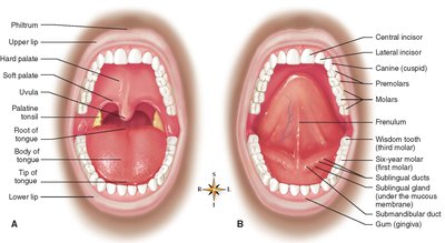
The mouth, or oral cavity, is the entry point for food and is involved in mechanical digestion and speech.
Roof: Hard palate (bone) and soft palate (muscle)
Floor: Formed by the tongue and its muscles
Uvula: Prevents food from entering nasal cavity; assists in swallowing
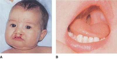
Tongue: Contains papillae and taste buds; anchored by lingual frenulum
Teeth
Teeth are essential for mechanical digestion.
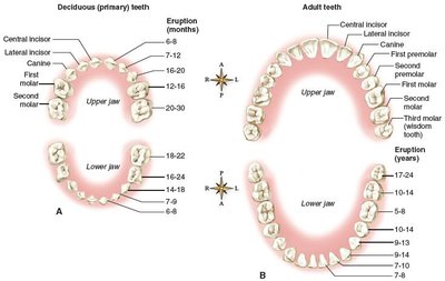
Types: Incisors, cuspids (canines), bicuspids (premolars), molars
Deciduous teeth: 20 primary teeth, erupt from 6 months to 2 years
Permanent teeth: 32 in most adults, erupt from 6 to 24 years
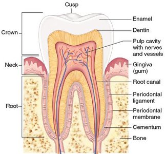
Structure: Crown (visible), neck (covered by gum), root (embedded in jaw)
Salivary Glands
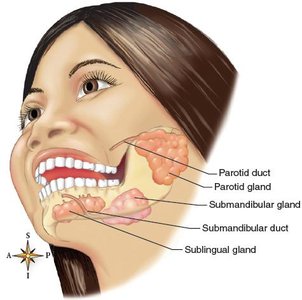
Salivary glands produce saliva, which contains enzymes for carbohydrate digestion.
Parotid: Largest, located near jaw
Submandibular: Beneath jaw
Sublingual: Under tongue
Salivary amylase: Begins starch digestion
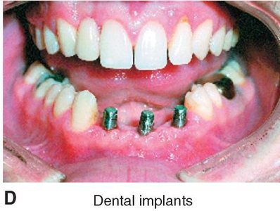
Disorders of the Mouth and Teeth
Common Conditions
Leukoplakia: Precancerous white patches
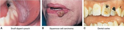
Snuff dipper’s pouch: Lesion from chewing tobacco
Squamous cell carcinoma: Most common oral cancer
Dental caries: Tooth decay leading to cavities
Gingivitis: Gum inflammation, often from poor hygiene
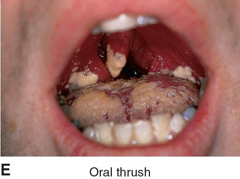
Thrush: Fungal infection (oral candidiasis)
Periodontitis: Inflammation of periodontal membrane, leading cause of adult tooth loss
Cleft lip/palate: Congenital defects from failed fusion during development
Pharynx and Esophagus
Pharynx
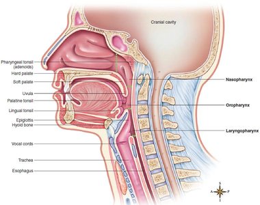
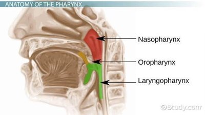
The pharynx is a muscular tube serving both respiratory and digestive functions. It is divided into three segments:
Nasopharynx
Oropharynx
Laryngopharynx
Esophagus
The esophagus is a muscular tube connecting the pharynx to the stomach.
Length: About 25 cm (10 inches)
Sphincters: Upper (UES) and lower (LES) esophageal sphincters regulate passage
Gastroesophageal Reflux Disease (GERD)
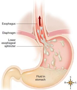
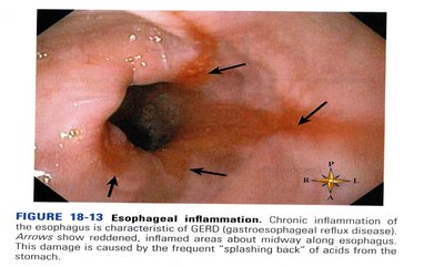
GERD is caused by backflow of acidic stomach contents into the esophagus, leading to heartburn and potential complications.
Symptoms: Heartburn, chest pain, asthma, esophageal irritation
Complications: Barrett esophagus, esophageal narrowing, bleeding
Hiatal Hernia
A hiatal hernia occurs when part of the stomach pushes through the diaphragm, often causing reflux symptoms. 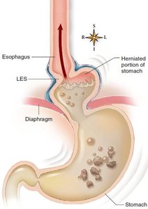
Stomach
Anatomy and Function
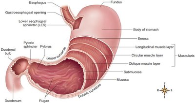
The stomach is a muscular pouch that mixes food with gastric juice, breaking it down into chyme.
Divisions: Fundus, body, pylorus
Pyloric sphincter: Regulates passage of food into duodenum
Rugae: Folds in mucosa when stomach is empty
Gastric glands: Secrete gastric juice and hydrochloric acid
Stomach Disorders
Pylorospasm: Spasms of pyloric sphincter, common in infants
Pyloric stenosis: Narrowing of pyloric opening
Ulcers: Open wounds caused by acid, often due to Helicobacter pylori infection or NSAIDs
Stomach cancer: Associated with alcohol, preserved foods, tobacco, and H. pylori
Small Intestine
Structure and Function
The small intestine is the primary site for digestion and absorption.
Sections: Duodenum, jejunum, ileum
Features: Circular folds (plicae), villi, and intestinal glands increase surface area
Disorders
Enteritis: Inflammation of the intestine
Gastroenteritis: Inflammation of stomach and intestines
Malabsorption syndrome: Failure to absorb nutrients properly
Maldigestion: Deficit of digestive enzymes or bile salts
Liver, Gallbladder, and Pancreas
Liver
The liver is the largest gland, classified as an exocrine gland, and produces bile for fat digestion.
Bile ducts: Hepatic, cystic, and common bile ducts
Gallbladder
The gallbladder stores and concentrates bile.
Disorders
Gallstones (cholelithiasis): Crystallized bile pigments and calcium salts
Cholecystitis: Inflammation of gallbladder
Hepatitis: Liver inflammation from toxins, bacteria, viruses
Cirrhosis: Degeneration of liver tissue, often from chronic alcohol abuse
Portal hypertension: High blood pressure in hepatic portal veins
Pancreas
The pancreas produces digestive enzymes and hormones (insulin, glucagon).
Pancreatitis: Inflammation, often from blocked ducts
Cystic fibrosis: Thick secretions block pancreatic juice
Pancreatic cancer: Highly fatal
Large Intestine, Appendix, and Peritoneum
Large Intestine
The large intestine absorbs water and forms feces.
Divisions: Cecum, colon (ascending, transverse, descending, sigmoid), rectum, anal canal
Conditions: Diarrhea, constipation, diverticulitis, colitis, colorectal cancer
Appendix
The vermiform appendix is a blind tube attached to the cecum; inflammation (appendicitis) is a common surgical emergency.
Peritoneum
The peritoneum is a large serous membrane lining the abdominal cavity and covering organs.
Extensions: Mesentery and greater omentum
Conditions: Peritonitis (inflammation), ascites (fluid accumulation)
Digestion and Absorption
Mechanical and Chemical Digestion
Mechanical digestion: Chewing, swallowing, peristalsis, segmentation
Chemical digestion: Enzymatic breakdown of food molecules
Enzymes and Hydrolysis
Enzymes: Protein catalysts for specific reactions
Hydrolysis: Addition of water to break down molecules
Carbohydrate Digestion
Pancreatic amylase: Starch to maltose
Maltase, sucrase, lactase: Disaccharides to glucose
Protein Digestion
Pepsin: Partial digestion in stomach
Trypsin: Complete digestion in small intestine
Peptidases: Final breakdown to amino acids
Fat Digestion
Bile: Emulsifies fats
Pancreatic lipase: Fats to fatty acids and glycerol
Absorption
Site: Small intestine (most nutrients and water), large intestine (some water)
Adaptations: Villi and microvilli increase surface area
Summary Table: Digestive Enzymes and Their Functions
Enzyme | Source | Substrate | End Product |
|---|---|---|---|
Salivary amylase | Salivary glands | Starch | Maltose |
Pepsin | Stomach | Proteins | Peptides |
Trypsin | Pancreas | Peptides | Amino acids |
Lipase | Pancreas | Fats | Fatty acids, glycerol |
Maltase, sucrase, lactase | Intestinal glands | Disaccharides | Glucose |
Key Equations
Hydrolysis Reaction
General equation for hydrolysis of a disaccharide:
Clinical Applications
Malocclusion
Malocclusion refers to misalignment of teeth, which can cause pain and functional problems. Orthodontics is the branch of dentistry that addresses these issues. 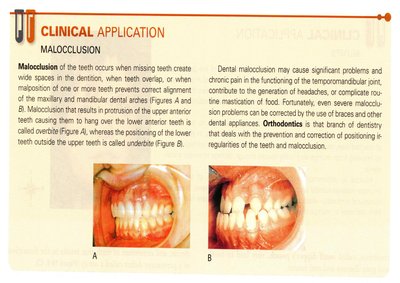
Mumps
Mumps is a viral disease affecting the parotid salivary glands, leading to swelling and potential complications. 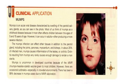
Review Questions
Which organ is considered an accessory organ of the digestive system? Answer: Teeth
Which disorder of the mouth is caused by a yeastlike fungal organism? Answer: Candidiasis (Thrush)
In an infant, when the pyloric fibers do not relax, food is unable to leave the stomach. This condition is called: Answer: Pylorospasm
What is the general term that refers to a group of symptoms resulting from the failure of the small intestines to absorb nutrients properly? Answer: Malabsorption syndrome
Hepatitis, chronic alcohol abuse, malnutrition, or infection may lead to a degenerative liver condition known as: Answer: Cirrhosis
Which type of autoimmune colitis often affects the small intestine? Answer: Crohn disease
What are the main processes of mechanical digestion? Answer: Chewing, swallowing, peristalsis
