 Back
BackDigestive System: Structure, Function, and Processes
Study Guide - Smart Notes
 Tailored notes based on your materials, expanded with key definitions, examples, and context.
Digestive System Overview
Introduction to the Digestive System
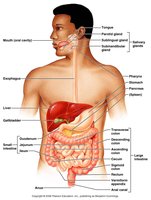
The digestive system is responsible for breaking down food into absorbable units to provide nutrients for cellular activity. It consists of the alimentary canal and accessory organs that aid in both mechanical and chemical digestion.
Alimentary canal: Mouth, pharynx, esophagus, stomach, small intestine, large intestine
Accessory organs: Salivary glands, teeth, tongue, liver, gallbladder, pancreas
Structure of Alimentary Canal Organs
Four Tunics of the Alimentary Canal
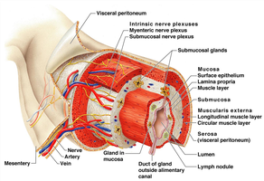
The walls of the alimentary canal are composed of four basic layers, each with specialized functions:
Mucosa: Innermost layer; consists of surface epithelium, lamina propria, and muscularis mucosae. Functions in secretion, absorption, and protection.
Submucosa: Connective tissue containing blood vessels, nerves, and glands.
Muscularis externa: Responsible for segmentation and peristalsis; consists of circular and longitudinal muscle layers.
Serosa: Outermost layer; also known as the visceral peritoneum.
Organs of the Digestive System
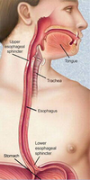
Mouth, Pharynx, and Esophagus
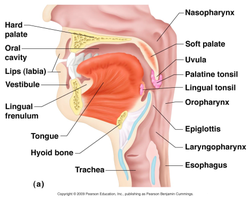
Mouth/Oral cavity: Site of ingestion, mechanical breakdown (mastication), and mixing with saliva.
Pharynx: Passageway for air and food; food is propelled to the esophagus by peristalsis, involving longitudinal and circular muscle layers.
Esophagus: Muscular tube (~10 inches) conducting food from pharynx to stomach via peristalsis; passageway for food only.
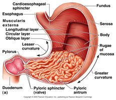
Stomach
Stomach Anatomy and Regions
The stomach is a muscular organ with several regions and specialized structures for digestion.
Cardiac region: Near the heart
Fundus: Expanded portion lateral to the cardiac region
Body: Main central region
Pylorus: Funnel-shaped terminal end leading to the duodenum
Muscularis externa: Includes an additional oblique muscle layer for churning food
Rugae: Folds in the mucosa that allow expansion
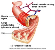
Small Intestine
Structural Modifications for Absorption
The small intestine is the primary site for nutrient absorption, with three key adaptations to increase surface area:
Circular folds (plicae circulares): Deep folds of mucosa and submucosa
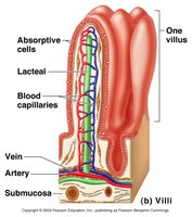
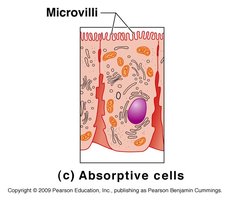
Villi: Fingerlike projections of the mucosa
Microvilli: Tiny projections on absorptive cells, forming a brush border
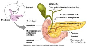
Relationship with Pancreas, Liver, and Gallbladder
The small intestine receives digestive enzymes from the pancreas and bile from the liver and gallbladder, essential for chemical digestion.
Pancreatic ducts deliver enzymes
Bile duct delivers bile for fat emulsification
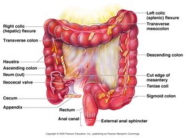
Large Intestine
Structure and Function
The large intestine absorbs water and electrolytes, forming and storing feces.
Cecum: Saclike first part
Appendix: Lymphatic tissue attached to the cecum
Colon: Ascending, transverse, descending, and sigmoid sections
Rectum and anal canal: Terminal portions
Digestive Processes
Physical (Mechanical) Digestion
Physical digestion involves breaking food into smaller pieces without chemical change to the food molecules.
Mastication: Chewing in the mouth
Churning: Mixing in the stomach
Segmentation: Rhythmic contractions in the small intestine
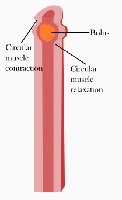
Peristalsis: Propels food along the tract
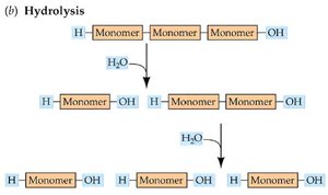
Chemical Digestion
Chemical digestion uses enzymes to break down complex molecules into absorbable units.
Enzymes are secreted by intrinsic and accessory glands into the digestive tract lumen.
Hydrolysis is the main chemical process, where water is used to break covalent bonds in food molecules.
Summary Tables: Chemical Digestion and Absorption
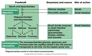
Carbohydrate Digestion and Absorption
Foodstuff | Enzyme(s) and Source | Site of Action |
|---|---|---|
Starch and disaccharides | Salivary amylase, Pancreatic amylase | Mouth, Small intestine |
Lactose, Maltose, Sucrose | Brush border enzymes (dextrinase, glucoamylase, lactase, maltase, sucrase) | Small intestine |
Monosaccharides (glucose, galactose, fructose) | Absorbed into capillary blood in villi | Small intestine |
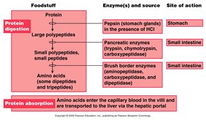
Protein Digestion and Absorption
Foodstuff | Enzyme(s) and Source | Site of Action |
|---|---|---|
Protein | Pepsin (stomach glands, with HCl) | Stomach |
Large polypeptides | Pancreatic enzymes (trypsin, chymotrypsin, carboxypeptidase) | Small intestine |
Small polypeptides, peptides | Brush border enzymes (aminopeptidase, carboxypeptidase, dipeptidase) | Small intestine |
Amino acids | Absorbed into capillary blood in villi | Small intestine |
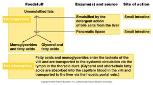
Fat Digestion and Absorption
Foodstuff | Enzyme(s) and Source | Site of Action |
|---|---|---|
Unemulsified fats | Bile salts (from liver), Pancreatic lipase | Small intestine |
Monoglycerides, fatty acids, glycerol | Absorbed into lacteals of villi | Small intestine |
Nucleic Acid Digestion
Enzymes: Pancreatic ribonuclease and deoxyribonuclease, brush border enzymes (nucleosidases, phosphatases)
Absorption: Pentose sugars, nitrogenous bases, and phosphate ions are absorbed into capillary blood in villi and transported to the liver via the hepatic portal vein.
Microscopic Anatomy of Digestive Organs
Histological Features
Esophagus: Stratified squamous epithelium; gastroesophageal junction marks transition to stomach epithelium.
Stomach: Gastric pits, parietal cells (secrete HCl), chief cells (secrete pepsinogen), mucosa, submucosa, muscularis mucosae.
Small intestine (Duodenum): Villi, Brunner’s glands (secrete alkaline mucus).
Small intestine (Ileum): Peyer’s patches (lymphatic tissue), villi.
Large intestine: Goblet cells (secrete mucus).
Liver: Central vein, hepatic portal triad (hepatic portal vein, hepatic artery, bile duct).
Pancreas: Pancreatic islets (endocrine), acinar cells (exocrine).
Additional info: Histological details are essential for understanding the specialized functions of each digestive organ, such as secretion, absorption, and immune defense.
