Back
BackEndocrine System: Adrenal Glands, Pancreas, and Hormonal Regulation
Study Guide - Smart Notes
 Tailored notes based on your materials, expanded with key definitions, examples, and context.
Adrenal Glands
Structure of the Adrenal Glands
The adrenal glands are pyramid-shaped organs located on the superior aspect of each kidney. Each gland consists of two main regions: the outer adrenal cortex and the inner adrenal medulla. The cortex is divided into three zones, each responsible for producing different steroid hormones.
Zona Glomerulosa: Produces mineralocorticoids (e.g., aldosterone).
Zona Fasciculata: Produces glucocorticoids (e.g., cortisol).
Zona Reticularis: Produces some glucocorticoids and androgenic steroids.
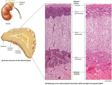
Additional info: All adrenal cortex hormones are derived from cholesterol and are classified as steroid hormones.
Hormones of the Adrenal Cortex
Mineralocorticoids: Aldosterone
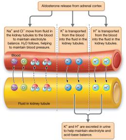
Aldosterone is the main mineralocorticoid, crucial for regulating sodium, potassium, and fluid balance.
Electrolyte Balance: Stimulates sodium and chloride reabsorption into the blood and potassium excretion into urine.
Fluid Volume: Indirectly increases water reabsorption by creating a concentration gradient, thus maintaining blood pressure.
Acid-Base Homeostasis: Promotes hydrogen ion excretion, helping maintain blood pH within 7.35–7.45.
Regulation: Aldosterone release is stimulated by high potassium, low pH, and angiotensin II. The hypothalamic-pituitary-adrenocortical (HPA) axis also plays a role via CRH and ACTH.
Disorders: Hyperaldosteronism can cause hypokalemia, hypernatremia, hypertension, and alkalosis.
Glucocorticoids: Cortisol
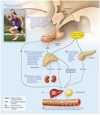
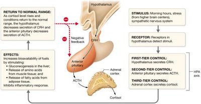
Cortisol is the primary glucocorticoid, essential for metabolic homeostasis and the stress response.
Metabolic Effects: Stimulates gluconeogenesis, increases blood glucose, mobilizes amino acids and fatty acids.
Anti-inflammatory Effects: Suppresses immune responses and inflammation.
Stress Response: Prepares the body for acute stress by mobilizing energy reserves and suppressing non-essential functions.
Regulation: Controlled by the HPA axis through a negative feedback loop involving CRH (hypothalamus), ACTH (anterior pituitary), and cortisol (adrenal cortex).
Disorders:
Hypercortisolism (Cushing's Syndrome/Disease): Excess cortisol causes fat redistribution, muscle wasting, hyperglycemia, hypertension, and increased infection risk.
Adrenal Insufficiency (Addison Disease): Deficiency of cortisol and aldosterone leads to fluid, electrolyte, and acid-base imbalances, and risk of adrenal crisis.

Androgenic Steroids
Produced in small amounts by the adrenal cortex, these hormones can be converted to testosterone or estrogen in the circulation and influence reproductive organs and other tissues.
Hormones of the Adrenal Medulla
The adrenal medulla contains chromaffin cells that secrete catecholamines (epinephrine and norepinephrine) in response to sympathetic nervous system stimulation. These hormones mediate the body's immediate response to stress ("fight or flight").
Increase heart rate and contractility
Dilate bronchioles
Constrict blood vessels to skin and viscera, dilate vessels to skeletal muscle
Dilate pupils, decrease digestive and urinary functions
Regulation involves sympathetic stimulation and ACTH/cortisol influence.
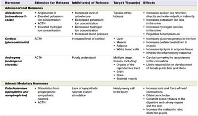
Summary Table: Hormones of the Adrenal Gland
Hormone | Stimulus for Release | Inhibitor(s) of Release | Target Tissues | Effects |
|---|---|---|---|---|
Aldosterone | Angiotensin-II, ACTH, elevated K+, decreased pH | Increased aldosterone, increased BP, increased Na+ | Kidney tubules | Increases Na+ retention, K+ and H+ excretion, maintains BP |
Cortisol | ACTH | Increased cortisol | Liver, muscle, adipose, WBCs | Stimulates gluconeogenesis, protein/fat breakdown, anti-inflammatory |
Androgens | ACTH | Poorly understood | Multiple targets | Sex characteristics, bone/muscle growth |
Catecholamines | Sympathetic stimulation, ACTH | Lack of stimulation | Nearly every cell | Fight-or-flight responses |
Pancreas and Glucose Homeostasis
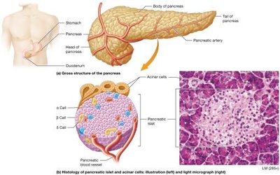
Structure of the Pancreas
The pancreas is a club-shaped organ with both endocrine and exocrine functions. Endocrine cells are organized into pancreatic islets, containing alpha (glucagon), beta (insulin), and delta (somatostatin) cells.
Hormones of the Endocrine Pancreas
Glucagon
Glucagon increases blood glucose by promoting glycogenolysis, gluconeogenesis, protein and fat breakdown, and ketone body formation. It is secreted in response to low blood glucose, sympathetic stimulation, and certain amino acids.
Excess ketone bodies can cause ketoacidosis, especially during starvation.
Insulin
Insulin lowers blood glucose by promoting uptake and storage of glucose, lipids, and amino acids. It is secreted in response to high blood glucose and is essential for cellular glucose uptake.
Hypoglycemia: Excess insulin causes dangerously low blood glucose.
Hyperglycemia: Insufficient insulin or resistance leads to high blood glucose (diabetes mellitus).
Type 1 Diabetes Mellitus: Autoimmune destruction of beta cells; requires insulin therapy.
Type 2 Diabetes Mellitus: Insulin resistance; often associated with obesity and may require lifestyle changes, oral hypoglycemics, or insulin.
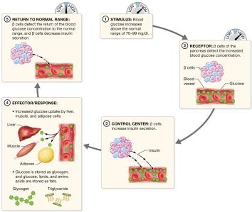
Blood Glucose Regulation
High Blood Glucose
Stimulus: Blood glucose rises above normal (70–99 mg/dL).
Receptor: Beta cells detect increased glucose.
Control Center: Beta cells secrete insulin.
Effector: Insulin increases glucose uptake and storage.
Return to Normal: Beta cells decrease insulin as glucose normalizes.
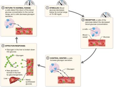
Low Blood Glucose
Stimulus: Blood glucose falls below normal.
Receptor: Alpha cells detect decreased glucose.
Control Center: Alpha cells secrete glucagon.
Effector: Glucagon increases glucose production and release.
Return to Normal: Alpha cells decrease glucagon as glucose normalizes.
Summary Table: Hormones of the Pancreas and Other Endocrine Organs
Organ/Tissue | Hormone(s) | Target Tissue(s) | Effects |
|---|---|---|---|
Pancreas | Insulin | Most cells | Stimulates glucose uptake, glycogenesis, lipogenesis |
Pancreas | Glucagon | Liver, muscle, adipose | Stimulates glycogenolysis, gluconeogenesis, ketogenesis |
Thymus | Thymosin, Thymopoietin | T lymphocytes | Promote T cell maturation |
Testes | Testosterone | Male reproductive organs, others | Androgenic and anabolic effects |
Ovaries | Estrogens, Progesterone | Female reproductive organs, others | Secondary sex characteristics, menstrual cycle, pregnancy support |
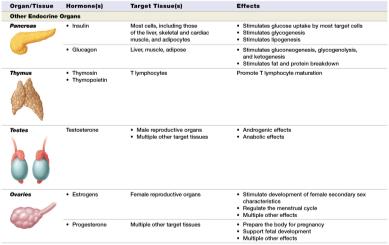
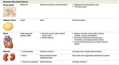
Hormones of Other Endocrine Organs and Tissues
Pineal Gland: Melatonin
Secretes melatonin, which regulates the sleep/wake cycle and promotes sleep, especially in response to darkness.
Adipose Tissue: Leptin
Secretes leptin, which acts on the brain to induce satiety and regulate feeding behavior. Its effectiveness in weight loss is limited due to complex regulatory mechanisms.
Heart: Atrial Natriuretic Peptide (ANP)
ANP is released in response to increased blood volume and stretch of the atria. It causes vasodilation and promotes sodium and water excretion, lowering blood pressure.
Kidneys: Erythropoietin, Renin, Calcitriol
Erythropoietin (EPO): Stimulates red blood cell production in response to hypoxia.
Renin: Initiates the renin-angiotensin-aldosterone system to regulate blood pressure.
Calcitriol: Active form of vitamin D, increases calcium absorption in the intestines.
Hormonal Control of Metabolic and Fluid Homeostasis
Metabolic Homeostasis
At rest, fasting: Thyroid hormones set basal metabolic rate; glucagon and growth hormone maintain blood glucose.
At rest, feeding: Insulin promotes nutrient storage; GH and IGF-1 stimulate protein synthesis; leptin and insulin promote satiety.
During exercise: Catecholamines and glucagon increase metabolic rate and fuel availability.
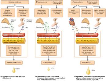
Fluid Homeostasis
Regulated by ADH, aldosterone, and ANP to maintain plasma volume and osmolarity.
Low plasma volume/high solute: Increased ADH and aldosterone, decreased urine output.
High plasma volume/low solute: Decreased ADH and aldosterone, increased ANP, increased urine output.
Hormonal Response to Stress
Stress triggers a coordinated hormonal response involving the hypothalamus, pituitary, adrenal glands, and other organs to maintain homeostasis and provide energy for the body.