 Back
BackEpithelial Tissue: Structure, Classification, and Function
Study Guide - Smart Notes
 Tailored notes based on your materials, expanded with key definitions, examples, and context.
Epithelial Tissue
Definition and Overview
Epithelial tissue is one of the four primary tissue types in the human body. It consists of closely packed cells that cover body surfaces, line internal cavities, and form glands. The main function of epithelial tissue is to provide protection, absorption, filtration, secretion, and sensory reception.
Tissue: A group of cells of similar structure performing a common function.
Special Characteristics of Epithelial Tissue
Epithelial tissues possess several unique features that distinguish them from other tissue types:
Cellularity: Composed almost entirely of cells with minimal extracellular matrix.
Polarity: Distinct apical (top) and basal (bottom) surfaces.
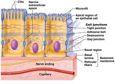
Cell Junctions: Specialized connections between adjacent cells, including tight junctions, adhesive belt junctions, desmosomes, and gap junctions.
Supported by Connective Tissue: The basal surface is attached to a basement membrane, which provides structural support.
Avascular but Innervated: Lacks blood vessels but contains nerve endings.
High Regenerative Capacity: Rapidly replaces damaged cells through mitosis.

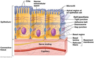
Lateral, Basal, and Apical Surface Features
The surfaces of epithelial cells have specialized features:
Lateral Surface: Contains cell junctions for adhesion and communication.
Basal Surface: Features the basal lamina and reticular fibers, forming the basement membrane. Functions as a selective filter and scaffolding for cell migration.
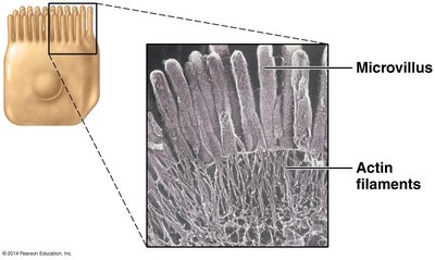
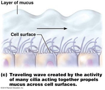
Apical Surface: May have microvilli (increase surface area for absorption) or cilia (motile structures that move substances across the surface).
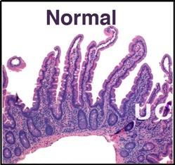
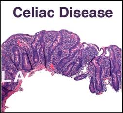
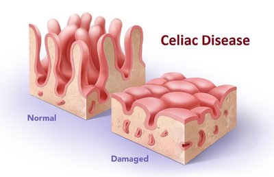
Clinical Example: Celiac Disease
Celiac disease is a genetic autoimmune disorder where the body attacks and destroys the villi and microvilli of the small intestine in response to gluten, resulting in poor nutrient absorption.
Normal villi: Intact microvilli increase absorption.
Celiac disease: Damaged villi and microvilli reduce absorption.
Functions of Epithelial Tissue
Epithelial tissues serve various functions depending on their location and structure:
Protection: Skin (epidermis)
Secretion: Glands
Absorption: Small intestine
Diffusion: Lungs
Filtration: Kidney
Sensory Reception: Nasal cavity (smell)
Classification and Naming of Epithelial Tissue
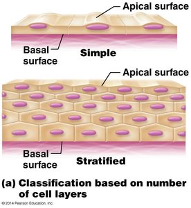
Classification Based on Layers
Epithelial tissues are classified by the number of cell layers:
Simple Epithelium: One cell layer; functions in absorption, secretion, and filtration.
Stratified Epithelium: Two or more cell layers; functions in protection.
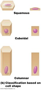
Classification Based on Cell Shape
The shape of the cells at the apical surface determines the second part of the name:
Squamous: Flat, scale-like cells
Cuboidal: Cube-shaped cells
Columnar: Tall, column-like cells
Types of Epithelial Tissue
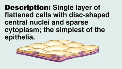
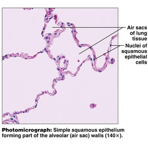
Simple Squamous Epithelium
Single layer of flattened cells; allows for diffusion and filtration.
Location: Lungs (air sacs), kidney (filtration), serous membranes (secretion)
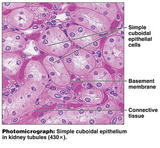
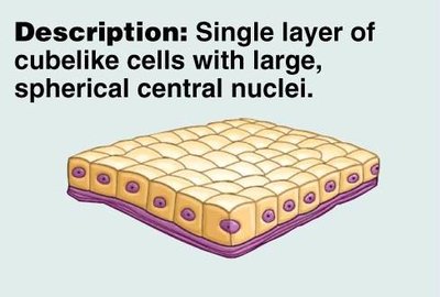
Simple Cuboidal Epithelium
Single layer of cube-shaped cells; specialized for absorption and secretion.
Location: Kidney tubules, some glands
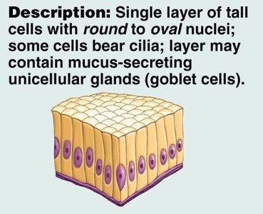
Simple Columnar Epithelium
Single layer of tall cells; may have cilia or microvilli. Functions in absorption, movement, and secretion.
Location: GI tract (absorption), small bronchi (movement of mucus), uterine tubes (propel ovum), secretion of mucus
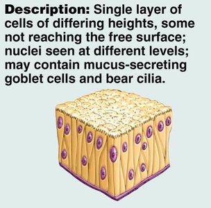
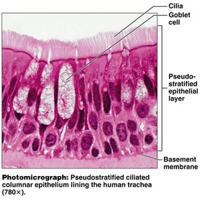
Pseudostratified Columnar Epithelium
Single layer of cells of varying heights; appears stratified but all cells touch the basement membrane. Functions in secretion and movement of mucus.
Location: Respiratory tract; usually ciliated
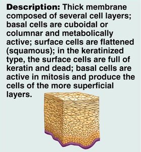
Stratified Epithelia
Composed of two or more layers; basal layer is germinating, apical layer is oldest. Provides protection.
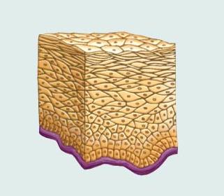
Stratified Squamous Epithelium
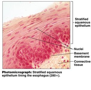
Multiple cell layers; apical layer is flat. May be keratinized (skin) or nonkeratinized (oral cavity, esophagus).
Keratinized: Contains tough protein keratin for protection.
Nonkeratinized: Moist surfaces, protection without keratin.
Stratified Cuboidal and Columnar Epithelium
Stratified cuboidal: Two layers of cube-shaped cells; protection and secretion (salivary glands). Stratified columnar: Several layers, apical layer tall; protection and secretion (male urethra, ducts of some glands).
Transitional Epithelium
Multiple layers that change shape when stretched; unique to urinary system, allows distention when structures fill with urine (lines urinary bladder).
Membranous vs Glandular Epithelium
Membranous Epithelium
Covers body surfaces and lines cavities; primary focus in anatomy and physiology.
Glandular Epithelium
Specialized for secretion; forms glands.
Endocrine glands: Lack ducts, secrete hormones into blood.
Exocrine glands: Secrete via ducts onto body surfaces or into cavities.
Types of Exocrine Glands
Unicellular: Goblet cells; produce mucus, found in respiratory and digestive tracts.
Multicellular: Classified by duct structure (simple = unbranched, compound = branched) and shape (tubular = tube-shaped, alveolar = spherical).
Type | Structure | Function | Location |
|---|---|---|---|
Simple Squamous | Single layer, flat cells | Diffusion, filtration | Lungs, kidney |
Simple Cuboidal | Single layer, cube-shaped | Absorption, secretion | Kidney tubules, glands |
Simple Columnar | Single layer, tall cells | Absorption, movement, secretion | GI tract, bronchi, uterine tubes |
Pseudostratified Columnar | Single layer, varying heights | Secretion, movement | Respiratory tract |
Stratified Squamous | Multiple layers, flat apical cells | Protection | Skin, oral cavity, esophagus |
Stratified Cuboidal | Two layers, cube-shaped | Protection, secretion | Salivary glands |
Stratified Columnar | Several layers, tall apical cells | Protection, secretion | Male urethra, ducts |
Transitional | Multiple layers, shape changes | Distention | Urinary bladder |
Additional info: Epithelial tissue classification is essential for understanding its function in different organs. The presence of microvilli and cilia on the apical surface is directly related to absorption and movement, respectively. The basement membrane is crucial for anchoring epithelial cells and facilitating tissue repair.
