 Back
BackFundamentals of the Nervous System and Nervous Tissue: Structured Study Notes
Study Guide - Smart Notes
 Tailored notes based on your materials, expanded with key definitions, examples, and context.
Fundamentals of the Nervous System and Nervous Tissue
Overview of the Nervous System
The nervous system is the primary controlling and communicating system of the body. It operates through rapid and specific electrical and chemical signals, enabling immediate responses to internal and external stimuli.
Sensory Input: Sensory receptors detect stimuli and send information to the CNS via afferent pathways.
Integration: The CNS processes and interprets sensory input, deciding on an appropriate response.
Motor Output: The CNS sends signals via efferent pathways to effectors (muscles and glands) to elicit a response.
Example: Seeing a red light (sensory input), integrating its meaning (stop), and pressing the brake (motor output).
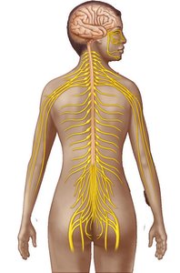
Additional info: The nervous system is highly integrated, with central and peripheral divisions working together to maintain homeostasis.
Structural and Functional Divisions of the Nervous System
Central Nervous System (CNS): Composed of the brain and spinal cord; serves as the integration and control center.
Peripheral Nervous System (PNS): Consists of nerves and ganglia outside the CNS; links the body to the CNS.
Functional Subdivisions of the PNS:
Sensory (Afferent) Division: Carries information from sensory receptors to the CNS.
Motor (Efferent) Division: Transmits signals from the CNS to effectors.
Somatic Nervous System: Controls voluntary movements of skeletal muscles.
Autonomic Nervous System (ANS): Regulates involuntary functions (smooth muscle, cardiac muscle, glands); includes sympathetic and parasympathetic divisions.

Neuroglia: Supporting Cells of the Nervous System
Types and Functions of Neuroglia
Neuroglia (glial cells) are specialized supporting cells that maintain and protect neurons. There are six types: four in the CNS and two in the PNS.
Astrocytes (CNS): Most abundant; support neurons, anchor them to capillaries, regulate chemical environment, and participate in information processing.
Microglial Cells (CNS): Monitor neuron health; act as macrophages to remove debris and pathogens.
Ependymal Cells (CNS): Line CNS cavities; circulate cerebrospinal fluid.
Oligodendrocytes (CNS): Form myelin sheaths around CNS axons.
Satellite Cells (PNS): Surround neuron cell bodies in the PNS; similar function to astrocytes.
Schwann Cells (PNS): Form myelin sheaths around PNS axons; vital for nerve regeneration.
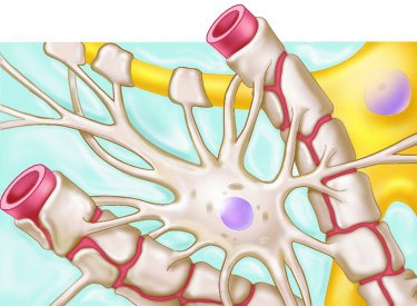
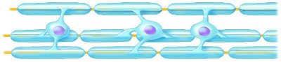
Additional info: Neuroglia are essential for maintaining the environment necessary for neuron function and for repairing nervous tissue.
Neurons: Structural Units of the Nervous System
Characteristics and Components of Neurons
Neurons are excitable cells that transmit electrical signals. They possess extreme longevity, are amitotic (do not divide), and have high metabolic rates requiring constant oxygen and glucose.
Cell Body (Soma): Contains nucleus, biosynthetic machinery, cytoskeletal elements, and pigment inclusions.
Dendrites: Short, branching processes; main receptive regions; convey graded potentials toward the cell body.
Axon: Single, long process; conducting region; generates and transmits action potentials away from the cell body.
Axon Terminals: Secretory region; release neurotransmitters to communicate with other cells.
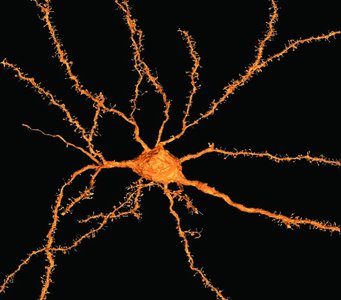
Additional info: Neurons depend on efficient transport mechanisms for distributing proteins and organelles along their axons.
Axonal Transport
Axonal transport is essential for moving materials between the cell body and axon terminals.
Anterograde Transport: Moves materials away from the cell body (e.g., mitochondria, enzymes).
Retrograde Transport: Moves materials toward the cell body (e.g., recycled organelles, signaling molecules).
Motor Proteins: Kinesin and dynein facilitate transport along microtubules.
Clinical Note: Some viruses and toxins exploit retrograde transport to reach the neuron cell body.
Myelin Sheath
The myelin sheath is a segmented, fatty covering that insulates axons and increases the speed of nerve impulse transmission.
Myelination in the PNS: Schwann cells wrap around axons, forming myelin sheaths; gaps between cells are called nodes of Ranvier.
Myelination in the CNS: Oligodendrocytes form myelin sheaths around multiple axons; no outer collar of perinuclear cytoplasm.
Nonmyelinated Axons: Thin axons are not myelinated and conduct impulses more slowly.
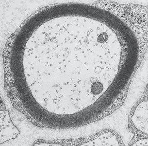
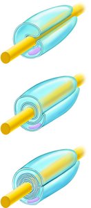
Additional info: Myelin sheaths are crucial for rapid communication in the nervous system.
Classification of Neurons
Structural Classification
Neurons are classified by the number of processes extending from the cell body:
Type | Structure | Location |
|---|---|---|
Multipolar | Three or more processes (one axon, multiple dendrites) | Most common; major type in CNS |
Bipolar | Two processes (one axon, one dendrite) | Special sense organs (retina, olfactory mucosa) |
Unipolar (Pseudounipolar) | Single process divides into peripheral and central branches | Found mainly in PNS ganglia; sensory neurons |
Additional info: Structural variations include Purkinje cells (cerebellum) and pyramidal cells (cerebral cortex).
Functional Classification
Neurons are also classified by the direction of impulse conduction:
Type | Function | Example |
|---|---|---|
Sensory (Afferent) | Transmit impulses toward CNS | Unipolar neurons in sensory ganglia |
Motor (Efferent) | Transmit impulses away from CNS to effectors | Multipolar neurons in CNS |
Interneurons | Integrate information within CNS | Multipolar neurons; Purkinje and pyramidal cells |
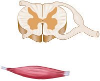
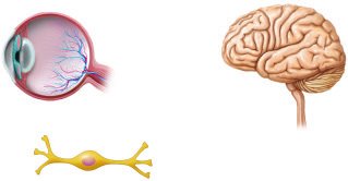
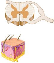
Additional info: Interneurons constitute over 99% of neurons and are essential for complex integration in the CNS.
Key Terminology: CNS vs. PNS
Term | Definition |
|---|---|
Nucleus | Collection of neuron cell bodies in the CNS |
Ganglion | Collection of neuron cell bodies in the PNS |
Tract | Bundle of axons in the CNS |
Nerve | Bundle of axons in the PNS |
Additional info: Understanding these terms is crucial for distinguishing anatomical structures in neurobiology.
