 Back
BackCh. 11 Fundamentals of the Nervous System and Nervous Tissue
Study Guide - Smart Notes
 Tailored notes based on your materials, expanded with key definitions, examples, and context.
The Nervous System: Overview and Functions
Major Functions of the Nervous System
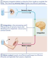
The nervous system is responsible for coordinating the body's responses to internal and external stimuli. It achieves this through three primary, overlapping functions:
Sensory Input: Sensory receptors detect changes inside or outside the body and relay this information to the central nervous system (CNS).
Integration: The CNS processes and interprets sensory input, determining the appropriate response.
Motor Output: The CNS sends signals to effectors (muscles or glands) to produce a response.
Divisions of the Nervous System
Anatomical and Functional Organization
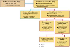
The nervous system is divided into two main anatomical parts, each with distinct roles:
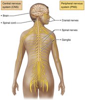
Central Nervous System (CNS): Composed of the brain and spinal cord, located in the dorsal body cavity. It serves as the integration and control center, interpreting sensory input and dictating motor output.
Peripheral Nervous System (PNS): Consists of nerves (cranial and spinal) and ganglia outside the CNS. It connects the CNS to limbs and organs, acting as a communication relay.
Functional Divisions of the PNS
The PNS is further divided based on function:
Sensory (Afferent) Division: Transmits sensory information from receptors to the CNS. Includes somatic sensory fibers (from skin, muscles, joints) and visceral sensory fibers (from organs).
Motor (Efferent) Division: Transmits commands from the CNS to effectors. Subdivided into:
Somatic Nervous System: Controls voluntary movements via skeletal muscles.
Autonomic Nervous System (ANS): Regulates involuntary functions (smooth muscle, cardiac muscle, glands). The ANS is further divided into:
Sympathetic Division: Mobilizes body systems during activity (fight or flight).
Parasympathetic Division: Conserves energy and promotes housekeeping functions during rest.
Neuroglia and Neurons
Cell Types in Nervous Tissue
Nervous tissue contains two principal cell types:
Neuroglia (Glial Cells): Support, protect, and insulate neurons. They do not conduct nerve impulses.
Neurons (Nerve Cells): Excitable cells that transmit electrical signals.
Neuroglia in the CNS
Astrocytes: Most abundant; support neurons, form the blood-brain barrier, guide neuron migration, and regulate the chemical environment.
Microglial Cells: Act as phagocytes, removing debris and pathogens.
Ependymal Cells: Line brain and spinal cord cavities; circulate cerebrospinal fluid (CSF).
Oligodendrocytes: Form myelin sheaths around CNS axons, increasing conduction speed.
Neuroglia in the PNS
Satellite Cells: Surround neuron cell bodies in ganglia; regulate the environment around neurons.
Schwann Cells: Form myelin sheaths around PNS axons; vital for nerve fiber regeneration.
Neurons: Structure and Function
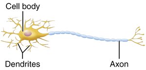
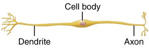
General Structure of a Neuron
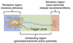
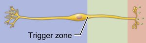
Neurons are highly specialized for communication. They have extreme longevity, are mostly amitotic, and have a high metabolic rate. Each neuron consists of a cell body (soma) and one or more processes (dendrites and axon).
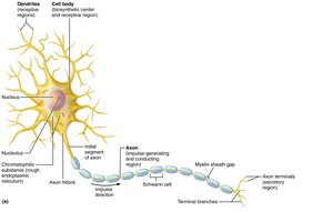
Neuron Processes: Dendrites and Axons
Dendrites: Short, branched processes that receive signals and convey them toward the cell body as graded potentials.
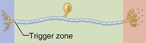
Axon: A single, long process that transmits impulses away from the cell body. Axons may branch and end in axon terminals, which release neurotransmitters.
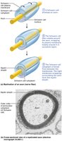
Myelin Sheath
The myelin sheath is a white, fatty covering that insulates axons and increases the speed of impulse transmission. Myelination differs between the PNS and CNS:
PNS: Schwann cells wrap around axons, forming the myelin sheath. Gaps between Schwann cells are called nodes of Ranvier.
CNS: Oligodendrocytes form myelin sheaths for multiple axons. No outer collar of perinuclear cytoplasm is present.
Classification of Neurons
Structural Classification
Neurons are classified by the number of processes extending from the cell body:
Multipolar Neurons: One axon and many dendrites; most common in the CNS.
Bipolar Neurons: One axon and one dendrite; found in special sensory organs (e.g., retina).
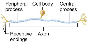
Unipolar (Pseudounipolar) Neurons: Single process that splits into two branches; mainly sensory neurons in the PNS.
Type | Structure | Location |
|---|---|---|
Multipolar | Many dendrites, one axon | CNS, motor neurons |
Bipolar | One dendrite, one axon | Retina, olfactory mucosa |
Unipolar | Single process, splits into two | Sensory neurons in PNS |
Functional Classification
Neurons are also classified by the direction in which they transmit impulses:
Sensory (Afferent) Neurons: Transmit impulses from sensory receptors to the CNS; mostly unipolar.
Motor (Efferent) Neurons: Carry impulses from the CNS to effectors; multipolar.
Interneurons (Association Neurons): Connect sensory and motor neurons within the CNS; most abundant type.
Type | Direction | Structure |
|---|---|---|
Sensory | To CNS | Unipolar |
Motor | From CNS | Multipolar |
Interneuron | Within CNS | Multipolar |
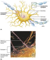
Synapses: Communication Between Neurons
Types and Structure of Synapses
Synapses are specialized junctions where neurons communicate with other neurons, muscle cells, or glands. There are two main types:
Electrical Synapses: Directly connect the cytoplasm of adjacent neurons via gap junctions, allowing rapid, bidirectional communication. Common in embryonic tissue and some brain regions.
Chemical Synapses: Use neurotransmitters to transmit signals across a synaptic cleft. Most common type in the nervous system.
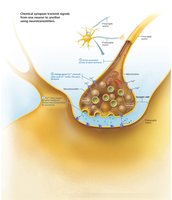
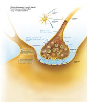
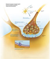
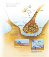
Chemical Synapse Transmission: Steps
Transmission at a chemical synapse involves six key steps:
An action potential arrives at the axon terminal of the presynaptic neuron.
Voltage-gated calcium channels open, and calcium enters the axon terminal.
Calcium entry causes synaptic vesicles to release neurotransmitter by exocytosis.
Neurotransmitter diffuses across the synaptic cleft and binds to receptors on the postsynaptic membrane.
Binding opens ion channels, creating graded potentials in the postsynaptic cell (can be excitatory or inhibitory).
Neurotransmitter effects are terminated by reuptake, enzymatic degradation, or diffusion away from the synapse.


Additional info:
Resting Membrane Potential: The resting membrane potential of a neuron is typically around -70 mV, generated by differences in ion concentrations and membrane permeability. The membrane is polarized, with the inside more negative than the outside.
Conduction Velocity: Action potentials (APs) travel faster in larger-diameter and myelinated axons. Myelinated axons use saltatory conduction, where the AP jumps between nodes of Ranvier, greatly increasing speed.
