 Back
BackFundamentals of the Nervous System and Nervous Tissue
Study Guide - Smart Notes
 Tailored notes based on your materials, expanded with key definitions, examples, and context.
The Nervous System: Overview
Introduction to the Nervous System
The nervous system is the master controlling and communicating system of the body. It enables rapid and specific communication between cells via electrical and chemical signals, resulting in almost immediate responses. The fundamental unit of communication is the nerve impulse (action potential).
Electrical signals: Transmit information quickly along neurons.
Chemical signals: Neurotransmitters released at synapses to communicate between neurons or between neurons and effectors.
Functions of the Nervous System
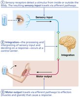
The nervous system performs three main, overlapping functions:
Sensory input: Gathering information from sensory receptors about internal and external changes.
Integration: Processing and interpreting sensory input and deciding on an appropriate response.
Motor output: Activating effectors (muscles and glands) to produce a response.
Organization of the Nervous System
Central and Peripheral Nervous Systems
The nervous system is divided into two principal parts:
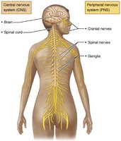
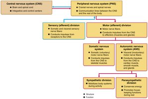
Central Nervous System (CNS): Consists of the brain and spinal cord, located in the dorsal body cavity. It serves as the integration and control center, interpreting sensory input and dictating motor output.
Peripheral Nervous System (PNS): Comprises nerves that extend from the brain and spinal cord, including cranial nerves, spinal nerves, and ganglia. The PNS connects the CNS to the rest of the body and includes the enteric nervous system in the gastrointestinal tract.
Functional Divisions of the PNS
The PNS is further divided functionally into:
Sensory (afferent) division: Transmits sensory information to the CNS.
Somatic sensory fibers: Carry impulses from skin, skeletal muscles, and joints.
Visceral sensory fibers: Carry impulses from visceral organs.
Motor (efferent) division: Transmits impulses from the CNS to effectors (muscles and glands).
Somatic nervous system: Controls voluntary movements of skeletal muscles.
Autonomic nervous system (ANS): Regulates involuntary activities of smooth muscle, cardiac muscle, and glands. The ANS is further divided into:
Sympathetic division: Mobilizes body systems during activity (fight or flight).
Parasympathetic division: Conserves energy and promotes housekeeping functions during rest.
Cells of Nervous Tissue
Neurons and Neuroglia
Nervous tissue consists of two principal cell types:
Neurons (nerve cells): Excitable cells that transmit electrical signals. They are the structural and functional units of the nervous system.
Neuroglia (glial cells): Support, protect, and insulate neurons. Types include astrocytes, oligodendrocytes, and Schwann cells.
Structure of a Motor Neuron
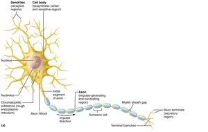
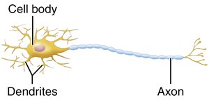
A typical neuron consists of a cell body (soma), dendrites, and an axon. The cell body contains the nucleus and organelles, dendrites receive signals, and the axon transmits impulses away from the cell body.
Neuroglia in the CNS
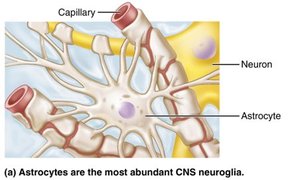
Astrocytes: Most abundant CNS neuroglia. They support and brace neurons, regulate exchanges between neurons and capillaries, control the chemical environment, and recycle neurotransmitters.
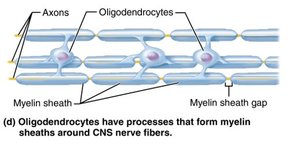
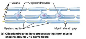
Oligodendrocytes: Form myelin sheaths around CNS nerve fibers, increasing the speed of impulse conduction.
Neuroglia in the PNS
Schwann cells: Form myelin sheaths around peripheral nerve fibers, vital for regeneration of damaged fibers and increasing conduction velocity.
Neuron Processes and Myelination
Myelin Sheath
The myelin sheath is a white, fatty substance that insulates many axons, especially long or large-diameter ones. Its main functions are to protect the axon and increase the speed of nerve impulse transmission. Myelinated axons conduct impulses much faster than nonmyelinated axons.
Nodes of Ranvier: Gaps between adjacent Schwann cells in the PNS or oligodendrocyte processes in the CNS. These are sites where axon collaterals can emerge and where action potentials are regenerated.
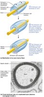
In the CNS, myelin is formed by oligodendrocytes, each of which can wrap up to 60 axons. In the PNS, Schwann cells form the myelin sheath by wrapping around a single axon segment.
Structure and Function of Neurons
Neuron Anatomy
Neurons have several key regions:
Dendrites: Short, branched processes that receive input from other neurons and convey it toward the cell body as graded potentials.
Cell body (soma): Contains the nucleus and is the site of most metabolic activities, including protein and neurotransmitter synthesis.
Axon: The conducting region that generates and transmits action potentials away from the cell body. The axon hillock is the trigger zone for action potential initiation.
Axon terminals: The secretory region where neurotransmitters are released to communicate with other cells.
Classification of Neurons
Structural Classification
Neurons are classified based on the number of processes extending from the cell body:
Multipolar neurons: One axon and two or more dendrites. Most common type in the CNS.
Bipolar neurons: One axon and one dendrite. Found in special sensory organs (e.g., retina, olfactory mucosa).
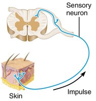
Unipolar (pseudounipolar) neurons: Single process that divides into two branches (peripheral and central processes). Most sensory neurons are unipolar.
Functional Classification
Sensory (afferent) neurons: Transmit impulses from sensory receptors toward the CNS. Almost all are unipolar.
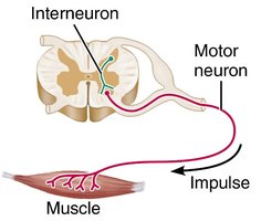
Motor (efferent) neurons: Carry impulses from the CNS to effectors (muscles and glands). Most are multipolar.
Interneurons: Lie between sensory and motor neurons within the CNS and are involved in integration. Most are multipolar.
Summary Table: Comparison of Structural Classes of Neurons
Neuron Type | Multipolar | Bipolar | Unipolar (Pseudounipolar) |
|---|---|---|---|
Processes | Many dendrites, one axon | One dendrite, one axon | One process that splits into two branches |
Location | CNS, motor neurons | Special sensory organs | Sensory neurons in PNS |
Example | Motor neuron | Retinal cell | Dorsal root ganglion cell |
Key Point: The structure of a neuron is closely related to its function in the nervous system.
