 Back
BackFundamentals of the Nervous System and Nervous Tissue: ANP Study Notes
Study Guide - Smart Notes
 Tailored notes based on your materials, expanded with key definitions, examples, and context.
Functions and Organization of the Nervous System
General Functions of the Nervous System
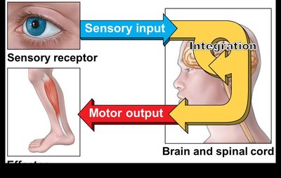
The nervous system is responsible for controlling and communicating information throughout the body. It has three main functions:
Sensory Input: Gathering information from sensory receptors to monitor changes (stimuli) inside and outside the body.
Integration: Processing and interpreting sensory input to determine if action is needed.
Motor Output: Activating muscles or glands in response to integrated stimuli.
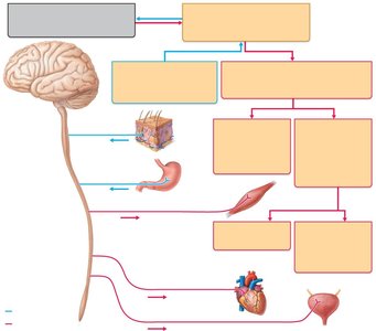
Organization of the Nervous System
Central Nervous System (CNS): Consists of the brain and spinal cord; serves as the integrative and control center.
Peripheral Nervous System (PNS): Includes cranial and spinal nerves; communication lines between the CNS and the rest of the body.
Sensory (Afferent) Division: Conducts impulses from receptors to the CNS; includes somatic and visceral sensory fibers.
Motor (Efferent) Division: Conducts impulses from the CNS to effectors (muscles and glands); includes the somatic (voluntary) and autonomic (involuntary) nervous systems.
Autonomic Nervous System (ANS): Subdivided into sympathetic (mobilizes body systems during activity) and parasympathetic (conserves energy, promotes housekeeping during rest) divisions.
Nervous Tissue: Support Cells (Neuroglia)
Neuroglia in the CNS
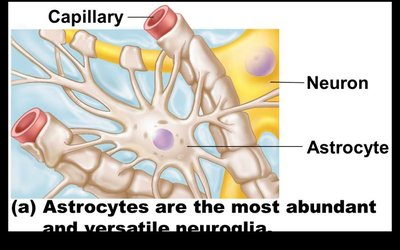
Neuroglia (glial cells) support, insulate, and protect neurons. There are four main types in the CNS:
Astrocytes: Abundant, star-shaped cells that brace neurons, form barriers between capillaries and neurons, and control the chemical environment of the brain.
Microglia: Spiderlike phagocytes that dispose of debris and defend CNS cells.
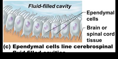
Ependymal Cells: Line cavities of the brain and spinal cord; cilia assist with circulation of cerebrospinal fluid.
Oligodendrocytes: Wrap around nerve fibers in the CNS and produce myelin sheaths.


Neuroglia in the PNS
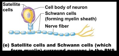
Satellite Cells: Protect neuron cell bodies in the PNS.
Schwann Cells: Form myelin sheaths around neurons in the PNS.
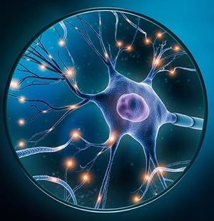
Neurons: Structure and Classification
General Structure of a Neuron
Neurons are the structural and functional units of the nervous system. They are highly specialized for impulse conduction, have extreme longevity, are mostly amitotic, and have a high metabolic rate.
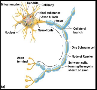
Cell Body (Soma/Perikaryon): Biosynthetic center containing the nucleus, nucleolus, and Nissl bodies (rough ER).
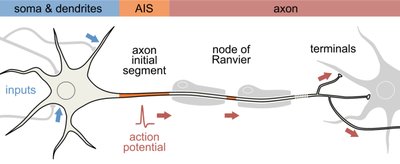
Processes: Armlike extensions from the cell body; include dendrites (receive signals) and axons (send signals).
Nuclei: Clusters of neuron cell bodies in the CNS.
Ganglia: Clusters of neuron cell bodies in the PNS.
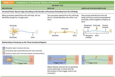
Structural Classification of Neurons
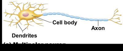
Multipolar Neurons: Many dendrites, one axon; most common, includes all motor and interneurons.
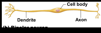
Bipolar Neurons: One dendrite and one axon; found in special sense organs (e.g., retina, olfactory mucosa).
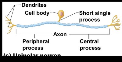
Unipolar Neurons: Single short process that splits into peripheral and central processes; mainly sensory neurons in PNS ganglia.
Neuron Type | Structure | Location |
|---|---|---|
Multipolar | Many dendrites, one axon | Motor neurons, interneurons |
Bipolar | One dendrite, one axon | Special senses (eye, nose) |
Unipolar | Single process splits into two | Sensory neurons in PNS |
Functional Classification of Neurons
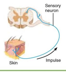
Sensory (Afferent) Neurons: Transmit impulses from sensory receptors toward the CNS; mostly unipolar.
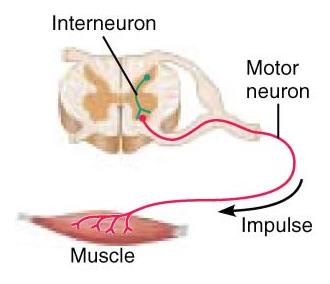
Motor (Efferent) Neurons: Carry impulses from the CNS to effectors; multipolar.
Interneurons (Association Neurons): Lie between sensory and motor neurons; shuttle signals through CNS pathways; most are multipolar and confined to the CNS.
Transmission of a Nerve Impulse: Membrane Potentials
Resting Membrane Potential
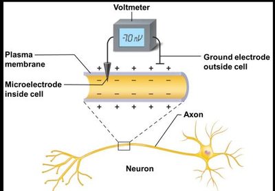
The resting membrane potential is the voltage difference across the membrane of a resting neuron, typically around –70 mV. The inside of the cell is more negative than the outside due to differences in ion concentrations and membrane permeability.
Generated by: Differences in ionic makeup (Na+ higher outside, K+ higher inside) and differential permeability of the plasma membrane.
Sodium-Potassium Pump: Maintains gradients by pumping 3 Na+ out and 2 K+ in.
Changes in Membrane Potential
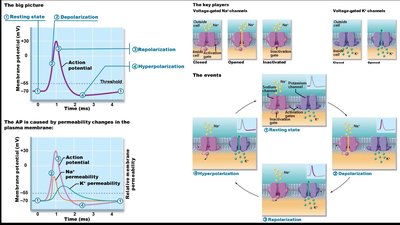
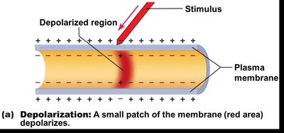
Depolarization: Membrane potential becomes less negative (toward zero); increases probability of nerve impulse.
Hyperpolarization: Membrane potential becomes more negative; reduces probability of nerve impulse.
Repolarization: Return to resting membrane potential, mainly via K+ efflux and Na+/K+ pump.
Action Potentials
Action potentials are long-distance electrical signals generated by changes in membrane potential. They are all-or-none events and propagate along the axon.
Threshold: Minimum depolarization needed to trigger an action potential (usually 15–20 mV above resting potential).
Phases: Depolarization (Na+ influx), repolarization (K+ efflux), hyperpolarization (excess K+ outflow), and return to resting state.
Propagation: Action potential moves down the axon, jumping between nodes of Ranvier in myelinated fibers (saltatory conduction).
Intensity and Rate of Transmission
All-or-None Principle: Once threshold is reached, the action potential cannot be reversed.
Stimulus Intensity: Determined by the frequency of action potentials, not their size.
Conduction Velocity: Depends on axon diameter (larger = faster) and degree of myelination (myelinated = faster).
Nerve Fiber Classification
Fiber Group | Diameter | Myelination | Speed | Location |
|---|---|---|---|---|
Group A | Large | Myelinated | 150 m/s | Somatic sensory/motor fibers |
Group B | Intermediate | Lightly myelinated | 15 m/s | Autonomic fibers |
Group C | Small | Unmyelinated | 1 m/s | Autonomic fibers |
Disorders Related to Myelination
Multiple Sclerosis: Autoimmune disease that destroys myelin sheaths, replaced by scleroses; disrupts conduction, affecting vision, speech, and movement.
Transmission at Synapses
The Synapse
Synapses are junctions that mediate information transfer from one neuron to another or to an effector cell. Transmission is electrical within the neuron and chemical between neurons.
Presynaptic Neuron: Conducts impulses toward the synapse.
Postsynaptic Neuron: Transmits impulses away from the synapse.
Types: Axodendritic, axosomatic, axoaxonal, dendrodendritic, somatodendritic.
Events at the Synapse
Action potential arrives at axon terminal.
Voltage-gated Ca2+ channels open; Ca2+ enters terminal.
Ca2+ triggers synaptic vesicles to release neurotransmitter into synaptic cleft.
Neurotransmitter binds to receptors on postsynaptic membrane, opening ion channels.
Graded potential is generated in postsynaptic neuron.
Neurotransmitter is removed by enzymatic degradation, reuptake, or diffusion.
Postsynaptic Potentials and Summation
Types of Postsynaptic Potentials
EPSP (Excitatory Postsynaptic Potential): Depolarization that brings the neuron closer to threshold; Na+ influx greater than K+ efflux.
IPSP (Inhibitory Postsynaptic Potential): Hyperpolarization that moves the neuron further from threshold; increased K+ or Cl– permeability.
Summation
EPSPs and IPSPs can summate (add together) to influence the postsynaptic neuron.
If EPSPs predominate and reach threshold, an action potential is generated.
Neurotransmitters
Major Neurotransmitters and Their Functions
Acetylcholine (Ach): Excitatory and inhibitory; found at neuromuscular junctions and ANS synapses. Decreased in Alzheimer's disease.
Norepinephrine (NE): Excitatory and inhibitory; found in ANS synapses. Increased by cocaine and amphetamines, leading to overstimulation.
Dopamine: Excitatory; controls mood, emotion, and muscle movement. Decreased in Parkinson's disease.
Serotonin: Inhibitory; regulates mood, anxiety, sleep, and appetite. Elevated in schizophrenia.
Endorphins: Inhibitory; reduce pain perception by binding to opioid receptors. Mimicked by morphine and heroin.
Developmental Aspects and Disorders of the Nervous System
Developmental Aspects
The nervous system forms during the first month of embryonic development.
Maternal infection and oxygen deprivation can cause severe damage.
Motor control development reflects myelination and maturation.
Brain growth ends in young adulthood; neurons die with age and are not replaced.
Disorders of the Nervous System
Neuroblastoma: Malignant tumor in children, usually in the PNS.
Rabies: Viral infection transmitted by animal bite; causes inflammation, delirium, and death.
Shingles: Reactivation of varicella-zoster virus; causes painful skin blisters and can recur.
