 Back
BackFundamentals of the Nervous System and Nervous Tissue – Study Notes
Study Guide - Smart Notes
 Tailored notes based on your materials, expanded with key definitions, examples, and context.
Fundamentals of the Nervous System and Nervous Tissue
Overview of the Nervous System
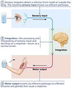
The nervous system is the master controlling and communicating system of the body. It uses electrical and chemical signals to coordinate rapid and specific responses to internal and external stimuli.
Sensory input: Information gathered by sensory receptors about changes inside and outside the body.
Integration: Processing and interpretation of sensory input to determine an appropriate response.
Motor output: Activation of effector organs (muscles and glands) to produce a response.
Divisions of the Nervous System
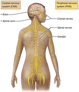
The nervous system is divided into two main parts:
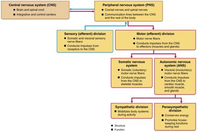
Central Nervous System (CNS): Consists of the brain and spinal cord. It is the integration and control center.
Peripheral Nervous System (PNS): Consists mainly of nerves that extend from the brain and spinal cord (cranial and spinal nerves) and ganglia. It serves as communication lines between the CNS and the rest of the body.
Functional Divisions of the PNS
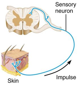
Sensory (afferent) division: Conducts impulses from receptors to the CNS. Includes somatic sensory fibers (from skin, muscles, joints) and visceral sensory fibers (from organs).
Motor (efferent) division: Conducts impulses from the CNS to effectors (muscles and glands). Subdivided into:
Somatic nervous system: Voluntary control of skeletal muscles.
Autonomic nervous system (ANS): Involuntary control of smooth muscle, cardiac muscle, and glands. Further divided into:
Sympathetic division: Mobilizes body systems during activity (fight or flight).
Parasympathetic division: Conserves energy and promotes housekeeping functions during rest.
Neuroglia (Glial Cells)
Neuroglia in the CNS
Neuroglia are supporting cells that protect, insulate, and support neurons. Four main types are found in the CNS:
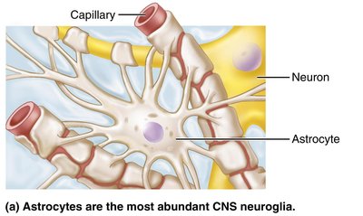
Astrocytes: Most abundant; support neurons, regulate the chemical environment, and help form the blood-brain barrier.
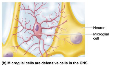
Microglial cells: Defensive cells that act as phagocytes, removing debris and pathogens.
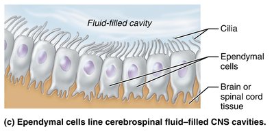
Ependymal cells: Line cerebrospinal fluid-filled cavities; cilia help circulate cerebrospinal fluid (CSF).
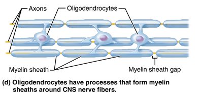
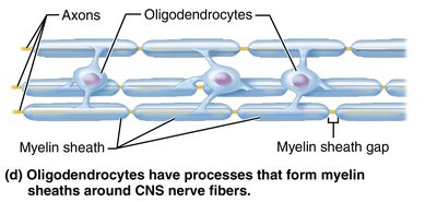
Oligodendrocytes: Form myelin sheaths around CNS nerve fibers, increasing conduction speed.
Neuroglia in the PNS
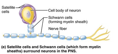
Satellite cells: Surround neuron cell bodies in the PNS; similar function to astrocytes.
Schwann cells: Form myelin sheaths around peripheral nerve fibers and assist in nerve regeneration.
Neurons (Nerve Cells)
Structure and Function
Neurons are the structural and functional units of the nervous system. They are specialized for conducting impulses and have extreme longevity, are mostly amitotic, and have a high metabolic rate.
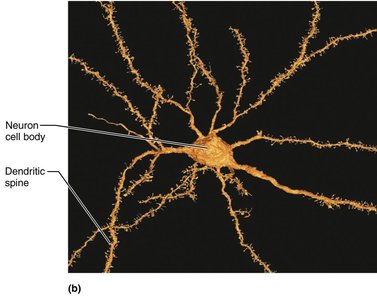
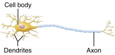
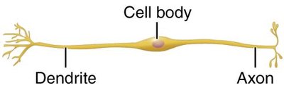
Cell body (soma): Contains the nucleus and organelles; biosynthetic and metabolic center.
Processes: Dendrites (receptive/input regions) and a single axon (conducting/output region).
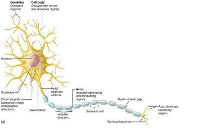
Dendrites
Short, branched extensions that receive signals and convey them toward the cell body as graded potentials.
Contain dendritic spines for increased surface area and synaptic input.
Axon
Each neuron has one axon, which may branch extensively at its end (axon terminals).
Conducts nerve impulses away from the cell body and releases neurotransmitters at axon terminals.
Axonal transport moves materials between the cell body and axon terminals (anterograde and retrograde transport).
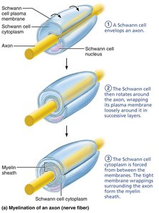
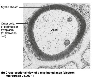
Myelin Sheath
The myelin sheath is a fatty, insulating layer that increases the speed of nerve impulse transmission.
In the PNS: Formed by Schwann cells wrapping around axons.
In the CNS: Formed by oligodendrocyte processes; one cell can myelinate multiple axons.
Classification of Neurons
Structural Classification
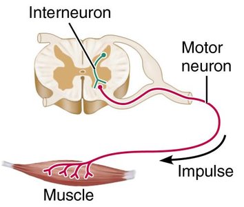
Multipolar: Many processes (1 axon, many dendrites); most common in CNS.
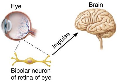
Bipolar: Two processes (1 axon, 1 dendrite); found in retina, ear, olfactory mucosa.
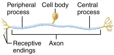
Unipolar (pseudounipolar): One process that splits into peripheral and central branches; mainly sensory neurons in PNS.
Type | Structure | Location |
|---|---|---|
Multipolar | Many dendrites, one axon | CNS, motor neurons |
Bipolar | One dendrite, one axon | Special senses (eye, ear, olfactory) |
Unipolar | Single process splits into two | Sensory neurons in PNS |
Functional Classification
Sensory (afferent) neurons: Transmit impulses from sensory receptors toward the CNS; mostly unipolar.
Motor (efferent) neurons: Carry impulses from the CNS to effectors; multipolar.
Interneurons: Lie between sensory and motor neurons; most abundant, found in CNS.
Membrane Potentials and Electrical Signals
Basic Principles of Electricity
Voltage (V): Potential energy generated by separated charges; measured in volts (V) or millivolts (mV).
Current (I): Flow of electrical charge (ions) between two points.
Resistance (R): Hindrance to charge flow.
Ohm’s Law:
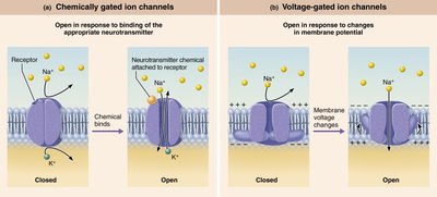
Ion Channels
Leakage (nongated) channels: Always open.
Gated channels: Open/close in response to stimuli (chemically, voltage, or mechanically gated).
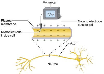
Resting Membrane Potential
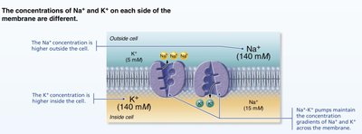
Neurons have a resting membrane potential (typically -70 mV), established by differences in ion concentrations and membrane permeability.
More K+ diffuses out than Na+ diffuses in, making the inside more negative.
The sodium-potassium pump ( out, in) maintains gradients.
Graded and Action Potentials
Graded Potentials
Short-lived, localized changes in membrane potential; essential for initiating action potentials.
Decay with distance; occur in dendrites and cell bodies.
Action Potentials (APs)
Brief reversal of membrane potential (~100 mV change).
Do not decay with distance; occur in axons.
All-or-none phenomenon: AP either happens completely or not at all.
Propagation and Conduction Velocity
APs propagate along axons by opening voltage-gated Na+ channels in adjacent regions.
Myelinated axons conduct impulses faster (saltatory conduction) than nonmyelinated (continuous conduction).
Synapses and Neurotransmitters
Synapses
Junctions that mediate information transfer between neurons or between a neuron and an effector cell.
Electrical synapses: Direct electrical coupling via gap junctions; rapid communication.
Chemical synapses: Use neurotransmitters to transmit signals across a synaptic cleft.
Postsynaptic Potentials
Excitatory postsynaptic potentials (EPSPs): Depolarize the postsynaptic membrane, increasing the likelihood of an AP.
Inhibitory postsynaptic potentials (IPSPs): Hyperpolarize the membrane, decreasing the likelihood of an AP.
Summation (temporal and spatial) integrates multiple inputs to determine if threshold is reached.
Neurotransmitters
Chemical Classification
Acetylcholine (ACh): Released at neuromuscular junctions; degraded by acetylcholinesterase.
Biogenic amines: Dopamine, norepinephrine, epinephrine, serotonin, histamine.
Amino acids: Glutamate, aspartate, glycine, GABA.
Peptides: Substance P, endorphins, somatostatin, CCK.
Purines: ATP, adenosine.
Gases and lipids: Nitric oxide, carbon monoxide, endocannabinoids.
Functional Classification
Excitatory vs. inhibitory: Effect depends on receptor type (e.g., ACh is excitatory at skeletal muscle, inhibitory at cardiac muscle).
Direct (fast) vs. indirect (slow): Direct neurotransmitters open ion channels; indirect act through second messengers (e.g., G protein pathways).
Neural Integration and Circuits
Patterns of Neural Processing
Serial processing: Input travels along one pathway to a specific destination (e.g., reflex arc).
Parallel processing: Input travels along several pathways, allowing complex responses.
Types of Neural Circuits
Diverging: One input, many outputs (amplifying circuit).
Converging: Many inputs, one output (concentrating circuit).
Reverberating: Signal travels through a chain of neurons, each feeding back to previous neurons.
Parallel after-discharge: Signal stimulates neurons arranged in parallel arrays.
Developmental Aspects of Neurons
Nervous system originates from the neural tube and neural crest (ectoderm).
Neuroblasts migrate, differentiate, and form synapses; astrocytes support synapse formation.
Many neurons die before birth if they fail to form synapses; apoptosis is common in development.
Neural plasticity (formation and removal of synapses) underlies learning and memory.
