 Back
BackFundamentals of the Nervous System and Nervous Tissue – Study Notes
Study Guide - Smart Notes
 Tailored notes based on your materials, expanded with key definitions, examples, and context.
Fundamentals of the Nervous System and Nervous Tissue
Introduction to the Nervous System
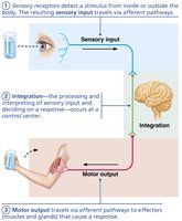
The nervous system is the master controlling and communicating system of the body. It uses electrical and chemical signals to coordinate rapid and specific responses to internal and external stimuli.
Sensory input: Information gathered by sensory receptors about changes inside and outside the body.
Integration: Processing and interpretation of sensory input, deciding what should be done at each moment.
Motor output: Activation of effector organs (muscles and glands) to produce a response.
Divisions of the Nervous System
The nervous system is divided into two main parts:
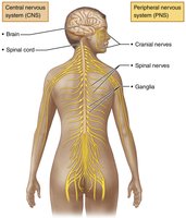
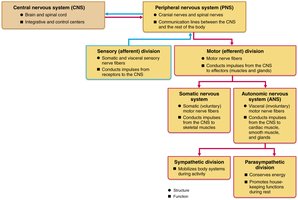
Central Nervous System (CNS): Consists of the brain and spinal cord. It is the integration and control center.
Peripheral Nervous System (PNS): Consists mainly of nerves that extend from the brain and spinal cord. It includes spinal nerves, cranial nerves, and the enteric nervous system (neurons in the gastrointestinal tract).
Functional Divisions of the PNS
Sensory (afferent) division: Carries impulses from receptors to the CNS. Includes somatic sensory fibers (from skin, muscles, joints) and visceral sensory fibers (from organs).
Motor (efferent) division: Transmits impulses from the CNS to effectors (muscles and glands). Subdivided into:
Somatic nervous system: Voluntary control of skeletal muscles.
Autonomic nervous system (ANS): Involuntary control of smooth muscle, cardiac muscle, and glands. Includes sympathetic (activates body systems) and parasympathetic (conserves energy) divisions.
Neuroglia (Glial Cells)
Neuroglia in the CNS
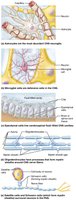
Neuroglia are supporting cells that protect, insulate, and support neurons. Four main types in the CNS:
Astrocytes: Most abundant; support neurons, regulate the chemical environment, and help form the blood-brain barrier.
Microglial cells: Act as phagocytes, removing debris and pathogens.
Ependymal cells: Line brain and spinal cord cavities; circulate cerebrospinal fluid (CSF).
Oligodendrocytes: Produce myelin sheaths in the CNS.
Neuroglia in the PNS
Satellite cells: Surround neuron cell bodies in the PNS; similar function to astrocytes.
Schwann cells: Form myelin sheaths around peripheral nerve fibers; vital for nerve regeneration.
Neurons (Nerve Cells)
Structure and Function
Neurons are the structural and functional units of the nervous system. They are specialized for conducting impulses and have the following characteristics:
Extreme longevity
Amitotic (do not divide after development)
High metabolic rate (require continuous oxygen and glucose)
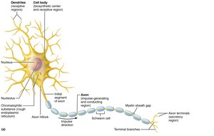
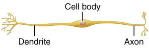
Each neuron has a cell body (soma) and one or more processes (dendrites and axons).
Neuron Processes
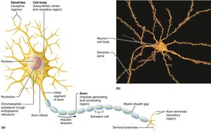
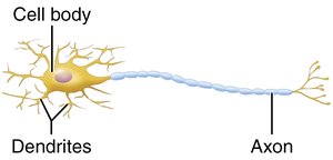
Dendrites: Short, branched processes that receive signals and convey them toward the cell body.
Axon: Long process that transmits impulses away from the cell body to other neurons or effectors. Axons may be myelinated or unmyelinated.
Myelin Sheath
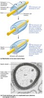
The myelin sheath is a fatty covering that insulates axons and increases the speed of impulse transmission.
In the PNS: Formed by Schwann cells; gaps between cells are called nodes of Ranvier.
In the CNS: Formed by oligodendrocytes; one cell can myelinate multiple axons.
Classification of Neurons
Structural Classification
Multipolar: Many processes (1 axon, many dendrites); most common in CNS.
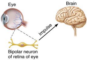
Bipolar: Two processes (1 axon, 1 dendrite); found in retina and olfactory mucosa.
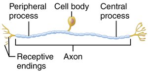
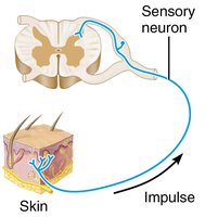
Unipolar: One process that splits into peripheral and central branches; mainly sensory neurons in PNS.
Type | Structure | Location |
|---|---|---|
Multipolar | 1 axon, many dendrites | CNS, motor neurons |
Bipolar | 1 axon, 1 dendrite | Retina, olfactory mucosa |
Unipolar | Single process | Sensory neurons in PNS |
Functional Classification
Sensory (afferent) neurons: Transmit impulses toward the CNS; mostly unipolar.
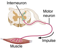
Motor (efferent) neurons: Carry impulses from the CNS to effectors; multipolar.
Interneurons: Connect sensory and motor neurons; most abundant, found in CNS.
Membrane Potentials
Basic Principles of Electricity
Voltage (V): Potential energy generated by separated charges.
Current (I): Flow of electrical charge between two points.
Resistance (R): Hindrance to charge flow.
Ohm’s Law:
Resting Membrane Potential
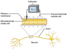
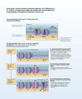
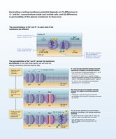
Neurons have a resting membrane potential (typically around -70 mV), maintained by differences in ion concentrations and membrane permeability. The sodium-potassium pump helps stabilize this potential by moving 3 Na+ out and 2 K+ in.
Changes in Membrane Potential
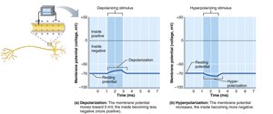
Depolarization: Membrane potential becomes less negative (closer to zero).
Hyperpolarization: Membrane potential becomes more negative.
Graded Potentials
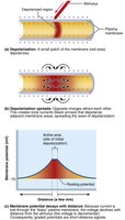
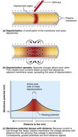
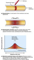
Graded potentials are short-lived, localized changes in membrane potential. They occur in dendrites and cell bodies and are essential for initiating action potentials.
Action Potentials
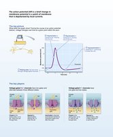
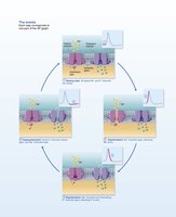
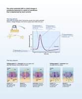
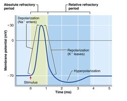
An action potential (AP) is a brief reversal of membrane potential that travels along the axon. It is an all-or-none event and does not decay with distance.
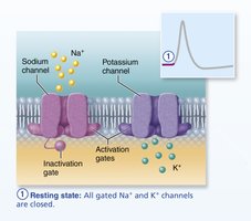
Resting state: All voltage-gated Na+ and K+ channels are closed.
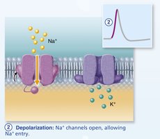
Depolarization: Na+ channels open, Na+ enters the cell.
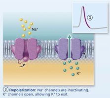
Repolarization: Na+ channels inactivate, K+ channels open, K+ exits the cell.
Hyperpolarization: Some K+ channels remain open, causing a slight dip below resting potential.
Propagation and Coding
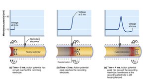
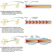
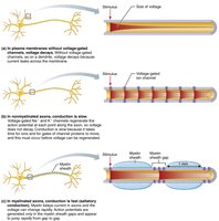
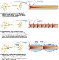
Action potentials propagate along axons by opening voltage-gated channels in adjacent segments.
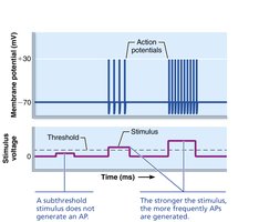
Stimulus intensity is coded by the frequency of action potentials, not their size.
Refractory Periods
Absolute refractory period: No new AP can be generated.
Relative refractory period: Only a strong stimulus can generate another AP.
Conduction Velocity
Depends on axon diameter (larger = faster) and degree of myelination (myelinated = faster, saltatory conduction).
Synapses
Types of Synapses
Electrical synapses: Direct electrical connection via gap junctions; rapid communication.
Chemical synapses: Most common; use neurotransmitters to transmit signals across a synaptic cleft.
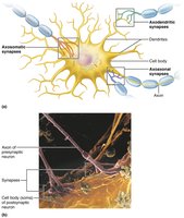
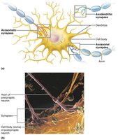
Chemical Synapse Transmission
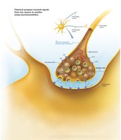
AP arrives at axon terminal.
Voltage-gated Ca2+ channels open; Ca2+ enters terminal.
Ca2+ entry causes neurotransmitter release by exocytosis.
Neurotransmitter diffuses across synaptic cleft and binds to receptors on postsynaptic membrane.
Binding opens ion channels, creating graded potentials.
Neurotransmitter effects are terminated by reuptake, degradation, or diffusion.
Postsynaptic Potentials
Excitatory postsynaptic potentials (EPSPs): Depolarize the postsynaptic membrane, increasing the likelihood of an AP.
Inhibitory postsynaptic potentials (IPSPs): Hyperpolarize the postsynaptic membrane, decreasing the likelihood of an AP.
Summation
Temporal summation: Multiple impulses from one neuron in rapid succession.
Spatial summation: Simultaneous impulses from multiple neurons.
Neurotransmitters
Chemical Classification
Acetylcholine (ACh): Released at neuromuscular junctions; degraded by acetylcholinesterase.
Biogenic amines: Dopamine, norepinephrine, epinephrine, serotonin, histamine; involved in emotion and biological clock.
Amino acids: Glutamate, aspartate, glycine, GABA.
Peptides: Substance P (pain), endorphins (natural opiates), gut-brain peptides.
Purines: ATP, adenosine.
Gases and lipids: Nitric oxide, carbon monoxide, endocannabinoids.
Functional Classification
Excitatory vs. Inhibitory: Depends on receptor type (e.g., ACh is excitatory at skeletal muscle, inhibitory at cardiac muscle).
Direct (fast) vs. Indirect (slow): Direct neurotransmitters open ion channels; indirect act through second messengers (e.g., G protein–coupled receptors).
Neural Integration
Patterns of Neural Processing
Serial processing: One pathway to a specific destination (e.g., reflex arc).
Parallel processing: Input travels along several pathways, allowing complex responses.
Neural Circuits
Diverging: One input, many outputs.
Converging: Many inputs, one output.
Reverberating: Signal travels through a chain of neurons, feeding back to previous neurons.
Parallel after-discharge: Signal stimulates neurons arranged in parallel arrays.
Developmental Aspects of Neurons
Nervous system originates from the neural tube and neural crest (ectoderm).
Neurons are amitotic after birth, except for olfactory neurons and those in the hippocampus.
Learning reinforces certain synapses and prunes others (neural plasticity).
