 Back
Fundamentals of the Nervous System and Nervous Tissue: Structured Study Notes
Study Guide - Smart Notes
 Tailored notes based on your materials, expanded with key definitions, examples, and context.
The Nervous System: Overview
Functions of the Nervous System
The nervous system is the body's primary communication and control system, responsible for sensing environmental changes, integrating information, and initiating responses. It consists of three main functions:
Sensory Input: Detects internal and external stimuli.
Integration: Processes and interprets sensory input.
Motor Output: Initiates responses by activating effector organs (muscles or glands).
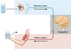
Basic Divisions of the Nervous System
Structural and Functional Organization
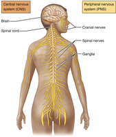
The nervous system is divided into structural and functional components:
Structural:
CNS (Central Nervous System): Brain and spinal cord.
PNS (Peripheral Nervous System): Peripheral nerves and ganglia.
Functional:
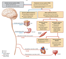
Sensory (Afferent) Division: Collects information and transmits it to the CNS.
Motor (Efferent) Division: Responds to information by transmitting signals from the CNS to effectors.
Further Functional Divisions
Sensory Division:
Somatic Sensory: Touch, pain, pressure, vibration, temperature, proprioception.
Visceral Sensory: Chemical changes and stretch in blood vessels and organs.
Motor Division:
Somatic Motor (SNS): Voluntary control of skeletal muscles.
Visceral Motor (ANS): Involuntary control of smooth muscle, cardiac muscle, and glands.
Nervous Tissue: Cellular Components
Types of Cells in Nervous Tissue
Nervous tissue is composed of two main cell types:
Neurons: Excitable cells that initiate and transmit nerve impulses. Adults have up to 100 billion neurons.
Neuroglial (Glial) Cells: Non-excitable cells that support and protect neurons. Adults have up to 1 trillion glial cells.
Characteristics of Neurons
Excitable: Responsive to stimulation.
Conductive: Electrical changes travel along the plasma membrane.
Secretory: Release neurotransmitters in response to electrical activity.
Extreme Longevity: Neurons can last a lifetime.
High Metabolic Rate: Require abundant nutrients and oxygen.
Non-mitotic: Most neurons do not divide, except neural stem cells in the hippocampus and olfactory bulb.
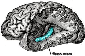
Additional info: Neural stem cells in the hippocampus are involved in memory processing.
Neuron Structure
Basic Structural Features
All neurons share certain structural features:
Dendrites: Conduct nerve impulses toward the cell body; receive input from other cells.
Soma (Cell Body): Control center; integrates and sends nerve impulses.
Axon: Conducts nerve impulses away from the cell body toward other cells.
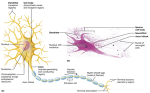
Dendrites
Short, branched processes that receive input and transfer it to the cell body.
The number of dendrites determines how many impulses a neuron can receive.
Soma (Cell Body)
Contains cytoplasm (perikaryon) and nucleus.
Serves as the control center and initiates electrical signals.
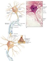
Axon
Long process that transmits nerve impulses away from the cell body.
Most neurons have only one axon.
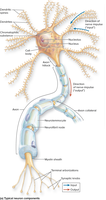
Information Flow in Neurons
Information flows from dendrites to the cell body, then to the axon, which stimulates a muscle or gland.
Classification of Neurons
Structural Classification
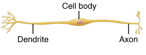
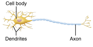
Neurons are classified by the number of processes emanating from the cell body:
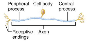
Unipolar: Single process; mostly sensory neurons.
Bipolar: Two processes (one axon, one dendrite); found in retina and olfactory epithelium.
Multipolar: Three or more processes (many dendrites, one axon); all motor neurons and interneurons.
Type | Processes | Location |
|---|---|---|
Unipolar | 1 | Sensory neurons |
Bipolar | 2 | Retina, olfactory epithelium |
Multipolar | >3 | Motor neurons, interneurons |
Functional Classification
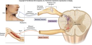
Sensory Neurons: Conduct sensory input; mostly unipolar; soma outside CNS.
Motor Neurons: Conduct motor output; multipolar; soma in CNS.
Interneurons: Facilitate communication between sensory and motor neurons; multipolar; entirely within CNS.
Synapses
Structure and Function
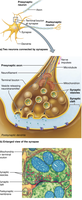
Synapses are sites where neurons communicate, usually via neurotransmitters. The presynaptic neuron sends information, and the postsynaptic neuron receives it. Most synapses are axodendritic, but some are axosomatic. Synaptic vesicles contain neurotransmitters, and the space between the synaptic knob and the effector is the synaptic cleft.
Glial Cells (Neuroglia)
Types and Functions
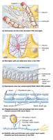
Glial cells occur in both CNS and PNS, are smaller than neurons, capable of mitosis, and do not transmit impulses. They physically protect, nourish, and support neurons, and outnumber neurons.
CNS Glial Cells:
Astrocytes: Most abundant; regulate neurotransmitter levels, blood flow, and ionic environment.
Microglia: Smallest, least abundant; immune cells.
Ependymal Cells: Line ventricles; circulate cerebrospinal fluid (CSF).
Oligodendrocytes: Produce myelin in CNS.
PNS Glial Cells:
Satellite Cells: Surround and nourish cell bodies in ganglia.
Schwann Cells (Neurolemmocytes): Produce myelin around peripheral axons.
Myelination of Axons
Process and Function
Myelination is the wrapping of axons with a myelin sheath, a protective fatty coating made from glial cell plasma membrane. Myelin insulates axons and speeds signal conduction (saltatory conduction). Oligodendrocytes myelinate CNS axons; Schwann cells myelinate PNS axons.
Unmyelinated Axons: Continuous conduction; slower.
Myelinated Axons: Saltatory conduction; faster and more energy-efficient.
Myelination in PNS vs CNS
PNS: Schwann cells myelinate a single axon.
CNS: Oligodendrocytes myelinate multiple axons.
Gross Anatomy of Nervous System
Gray and White Matter
Gray Matter: Neuronal cell bodies, dendrites, non-myelinated axons, neuroglia.
White Matter: Myelinated axons and neuroglia; called tracts in CNS, nerves in PNS.
Brain: Deep gray surrounded by white, with additional gray cortex on exterior.
Spinal Cord: Central gray surrounded by white; no cortex.
Nerves
Structure and Organization
Found in the PNS; contain both myelinated and nonmyelinated axons.
Wrapped in connective tissue:
Endoneurium: Around Schwann cell.
Perineurium: Around bundles of axons (fascicles).
Epineurium: Around the whole nerve.
Definitions:
Neuron: Nerve cell.
Nerve fiber: Long axon.
Nerve: Collection of axons in the PNS.
Neural Integration and Reflex Arcs
Integration Between CNS and PNS
PNS: Sensory and motor axons bundled in nerves; pathway from CNS to muscles, organs, glands.
CNS: Interneurons receive sensory info, transport it, and initiate responses.
Reflex Arc: Simplest example of neural integration.
Reflex Arcs
Monosynaptic Reflex: No interneuron; e.g., knee-jerk reflex.
Polysynaptic Reflex: Has interneuron; e.g., withdrawal reflex.
Neural Circuits
Types of Circuits
Diverging Circuits: One input to several outputs.
Converging Circuits: Several inputs to one output.
Reverberating Circuits: Neuronal feedback.
Processing:
Serial: One neuron to the next in a chain (reflex).
Parallel: One neuron has several perceptions (complex processing).
Disorders of Nervous Tissue
Multiple Sclerosis
Autoimmune disease causing demyelination of neurons.
Destruction of oligodendrocytes.
Impairs sensory perception and motor coordination.
Guillain-Barre Syndrome
Inflammation causes loss of myelin from peripheral nerves and spinal nerve roots.
Muscle weakness followed by paralysis; most people recover.
Regeneration of PNS Axons
Regeneration Process
PNS axons can regenerate if some neurilemma remains.
Depends on:
Amount of damage
Neurolemmocyte secretion of nerve growth factors
Distance between damaged axon and effector organ
Steps:
Axon end seals off
Schwann cells clean out dead axon
Axon filament grows through regeneration tube
Axon regenerates and new myelin sheath forms
In CNS, neuroglia do not form bands to guide regrowing axons, so regeneration is ineffective.
Additional info: Neural injuries in CNS tend to cause permanent dysfunction due to lack of regeneration.
